New Microscope Brings Glowing Cells into Focus
A New Tool for Studying Life in Motion
Advertisement


Researchers at Helmholtz Munich and the Technical University of Munich have developed a new microscope that significantly improves how bioluminescent signals in living cells can be observed. The system, known as QIScope, is built around a highly sensitive camera technology capable of detecting extremely low levels of light. With sharper image resolution, a wider field of view, and integration with other imaging methods, QIScope opens new opportunities for studying living systems in greater detail and over longer periods.
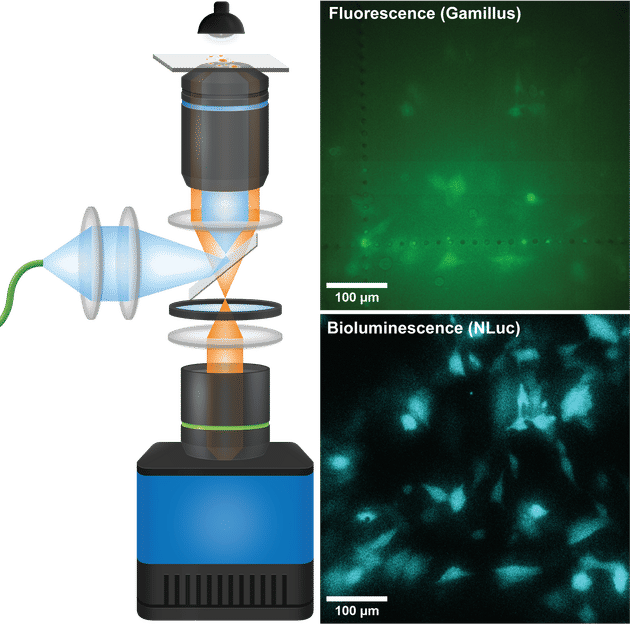
When imaging low protein levels in live cells on the high-sensitivity QIScope, bioluminescence (blue) significantly outperforms fluorescence (green)
Ruyu Ma - Helmholtz Munich
Bioluminescence: A Gentle Light for Biological Insight
Bioluminescence, the light emission produced by specific enzymes in certain living cells, is a powerful tool in life sciences. Unlike fluorescence imaging, which relies on strong external illumination that can interfere with cell behavior or obscure subtle signals, bioluminescence offers a gentler alternative for long-term observations. Its main drawback, however, is the faintness of the emitted light, which has made detailed imaging technically challenging.
Telescope-Inspired Design Meets Sensitive Sensor
To overcome this limitation, the team led by Dr. Jian Cui explored the use of quanta image sensors (QIS) – a new camera technology that they found outperforms the commonly used cameras (EMCCD) in low-light conditions. To harness the full potential of the QIS, the researchers developed an unconventional optical system that combines features of a telescope and a microscope, resulting in the creation of the QIScope. “To take full advantage of the sensor’s capabilities, we took inspiration from the optical layout of telescopes,” says Ruyu Ma, first author of the study and Doctoral Researcher at the Helmholtz Pioneer Campus. “By combining this approach with the QIS camera, we created a system that can reveal cellular processes with a clarity and sensitivity that was not possible with the state-of-the-art system.”
Capturing Subtle Changes in Living Cells
The team of researchers demonstrated that QIScope can track fine-scale dynamics in living cells – such as the movement of vesicles and the behavior of low-abundance proteins – over extended periods.
“Our microscope offers higher sensitivity, improved resolution, larger field of view, and higher dynamic range - all things you would want for challenging live-cell imaging experiments,” says study leader Jian Cui. “It also integrates other imaging methods such as epifluorescence and, in principle, phase contrast. This opens the door to observing living systems with much less disturbance, which is essential for understanding complex biological processes in health and disease.”
A New Tool for Studying Life in Motion
By addressing key limitations of traditional bioluminescence imaging, the QIScope provides researchers with a valuable tool for studying a range of biological systems – from single cells to organoids and tissue models. Its ability to reveal subtle and long-term changes in cell behavior may support progress in diverse research areas, including cell biology, disease modeling, and drug discovery.
Original publication
Ruyu Ma, Luciano M. Santino, Tomáš Chobola, Niklas Armbrust, Julian Geilenkeuser, Sapthagiri Sukumaran, Zhizi Jing, Anastasia Levkina, Korneel Ridderbeek, Tingying Peng, Dong-Jiunn Jeffery Truong, Sebastian Doll, Gil Gregor Westmeyer, Jian Cui; "A telescopic microscope equipped with a quanta image sensor for live-cell bioluminescence imaging"; Nature Methods, 2025-5-29


Other news from the department science


These products might interest you


Most read news


More news from our other portals


See the theme worlds for related content
Topic World Cell Analysis
Cell analyse advanced method allows us to explore and understand cells in their many facets. From single cell analysis to flow cytometry and imaging technology, cell analysis provides us with valuable insights into the structure, function and interaction of cells. Whether in medicine, biological research or pharmacology, cell analysis is revolutionizing our understanding of disease, development and treatment options.