New three-dimensional imaging method paves way for several research fields
Advertisement


A new method for 3D imaging and quantification of biological preparations ten times larger than the limit for the traditional confocal microscope is now being presented by researchers from Umeå University in Sweden in the journal Nature Methods.
Traditional biological imaging techniques are limited by several factors, such as the optical properties of the tissue and access to biological markers. A major challenge in this connection has been the creation of three-dimensional images of the expressions of specific genes and proteins in large biological preparations. It has been equally complicated to try to measure the mass/volume of cells or structures that express a specific protein in a specific organ, for instance.
The study now presented, under the direction of Associate Professor Ulf Ahlgren at the Umeå Center for Molecular Medicine (UCMM), in collaboration with Dr. James Sharpe at the Centre for Genomic Regulation in Barcelona and Professor Dan Holmberg at the Department of Medical Bioscience, Umeå University, describes an elaboration of the technology for optical projection tomography (OPT) that the two former researchers helped describe in the journal Science four years ago. The scientists have combined improvements in sample processing and tomographic data processing to develop a new method that make it possible to create 3D images of specifically dyed preparation that are one centimeter in size, of organs from adult mice and rats, for example.
Moreover, the authors describe how the new method can be used to automatically measure the number and volume of specifically dyed structures in large biological preparations. The technique requires no specially developed biological marker substances; instead, it makes use of antibodies that are in routine use in many research laboratories. The article exemplifies the potential of the technology by following the degradation of insulin-producing cells in intact pancreases from a mouse model for type-1 diabetes. In this case they demonstrate a direct connection between the volume of the remaining insulin-producing cells and the development of symptoms of diabetes.
The researchers project that it will be possible to use their method to address a great number of medical and biological issues. This may include such diverse fields as the formation of blood vessels in tumor models, the analysis of biopsies taken from patients (in cirrhosis of the liver, for instance), and autoimmune infiltration processes.


Other news from the department science


Most read news


More news from our other portals


See the theme worlds for related content
Topic world Fluorescence microscopy
Fluorescence microscopy has revolutionized life sciences, biotechnology and pharmaceuticals. With its ability to visualize specific molecules and structures in cells and tissues through fluorescent markers, it offers unique insights at the molecular and cellular level. With its high sensitivity and resolution, fluorescence microscopy facilitates the understanding of complex biological processes and drives innovation in therapy and diagnostics.
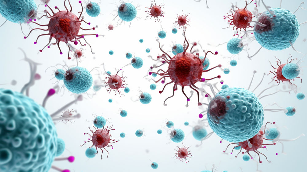
Topic world Antibodies
Antibodies are specialized molecules of our immune system that can specifically recognize and neutralize pathogens or foreign substances. Antibody research in biotech and pharma has recognized this natural defense potential and is working intensively to make it therapeutically useful. From monoclonal antibodies used against cancer or autoimmune diseases to antibody-drug conjugates that specifically transport drugs to disease cells - the possibilities are enormous