'Nanoshells' simultaneously detect and destroy cancerous cells
Advertisement


Researchers at rice University in Texas have developed a new approach to fighting cancer, based on nanoscale particles that can both detect and destroy cancerous cells. The report appears in the American Chemical Society's journal Nano Letters. Current molecular imaging approaches only detect the cancer but don't offer a method of treatment, according to the study's lead authors, Rebekah Drezek, Ph.D., and Jennifer West, Ph.D., both professors in the Department of Bioengineering at Rice.
"You can look for a molecular marker that may indicate a significant clinical problem, but you can't do anything about it [just through imaging]," says Drezek. "We don't want to simply find the cancerous cells. We would like to locate the cells, be able to make a rational choice about whether they need to be destroyed and, if so, proceed immediately to treatment."
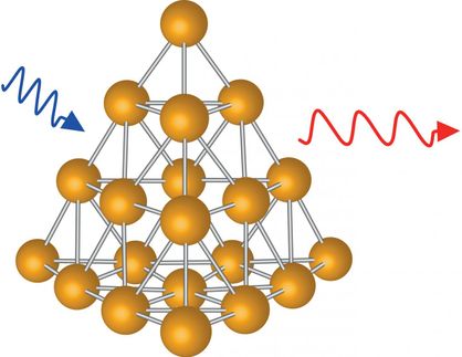
To this end, Drezek and West collaborated to develop a new imaging and treatment method based on metal "nanoshells" - tiny spheres of silica coated with a thin layer of gold. Nanoshells were invented by electrical engineer Naomi Halas, Ph.D., also of Rice University. Because these spheres are constructed on the nanometer scale, they exhibit unique size-dependent behavior, such as tunable optical properties. This allows researchers to design particles that scatter and absorb light at particular wavelengths.
The scattering of light provides the optical signal used to detect the cancer cells, which then "light up" when they come into contact with the nanoshells. In this study, the researchers designed the nanoshells to look for breast cancer biomarkers on the surface of the cancer cells. The technique can be readily extended to target other types of cancer or disease processes that have known surface markers.The additional ability of the particles to absorb light is used to generate heat, which then destroys the cancer cells. "Nanoshells are very unique in that we can engineer the particles so that both the optical scattering and absorption peaks occur in the near-infrared (NIR) spectral region where light penetration through tissue is highest," Drezek says. The NIR absorption also makes destruction of the targeted cells less invasive for patients because it uses a light source from outside the body that passes harmlessly through normal tissue and only heats the tissue containing nanoshells.
The new approach has some significant advantages over other alternatives that are under development, according to Drezek. For instance, optical imaging is much faster and less expensive than other medical imaging techniques. Gold nanoparticles are also more biocompatible than other types of optically active nanoparticles, such as quantum dots.
Gold is a chemically inert material that is well-known for its biocompatibility, which is why it has found use in a variety of medical applications in the past. "There is a prior history of the use of gold inside the body that makes the safety issues somewhat easier to address," Drezek says. Of course, any new technology requires extensive safety assessment before coming to market, but initial results from nanoshells testing are promising. Nanoshells developed for therapeutic applications have already been evaluated by Nanospectra Biosciences Inc., the Houston-based company that is commercializing the technology, with no ill effects found, according to Drezek.


Other news from the department science


