Sustainable Printing Inks for Functional Organs
The PhysioINK project is developing ink made from tissue proteins that can replicate physiological tissue structures without the need for chemical modification
Advertisement


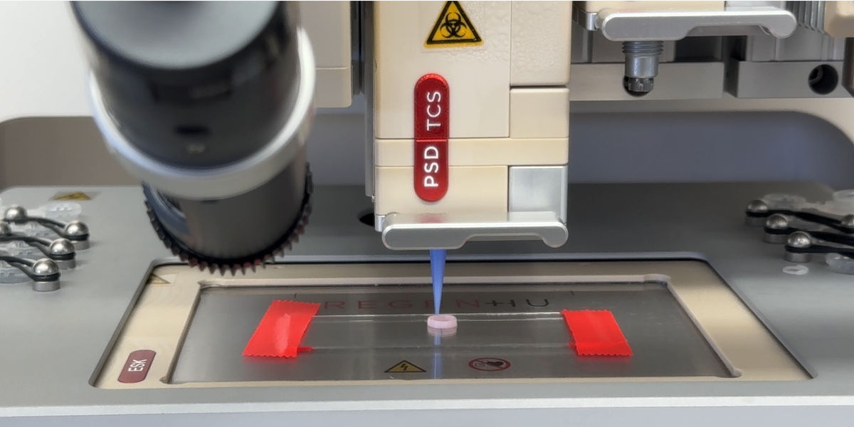
3D bioprinting opens up new possibilities for producing organs and tissues needed for applications such as organ transplants and drug development. However, while existing bioinks are cell-compatible and printable, they do not adequately replicate the physiological structure and composition of tissue. In the PhysioINK project, Fraunhofer researchers are developing bioinks from the most common tissue proteins to replicate physiological tissue structures without chemical modification.
The organ transplant waiting list numbers 8,000 people each year in Germany alone, and about ten percent of these never receive an organ. 3D bioprinting could help address the global shortage of donor organs by making it possible to automatically produce implantable organs or organ components tailored to individual patients “at the touch of a button.” This technology could also be used to print pathological tissue models that replicate diseased tissue, supporting disease research and drug development and testing. However, one challenge in 3D bioprinting remains unresolved: Bioinks either offer good printing properties but cannot replicate natural tissue structures, or they reproduce those structures but are not sufficiently printable. The Fraunhofer Institute for Silicate Research ISC, the Fraunhofer Institute for Applied Polymer Research IAP, the Fraunhofer Institute for Microstructure of Materials and Systems IMWS and the Bioanalytics and Bioprocesses branch of the Fraunhofer Institute for Cell Therapy and Immunology IZI are working to overcome this hurdle. In the PhysioINK project, they aim to develop sustainable, process-optimized bioinks that can precisely replicate physiological human organ structures and be used effectively in various 3D printing methods. The researchers plan to develop two applications: standardized intestinal tumor models for drug development and physiological cardiac tissue as a step toward functional implants for personalized regenerative medicine.
Highly concentrated printing inks based on collagen and elastin
“Until now, printing inks have been made from fully synthetic or heavily chemically modified materials. In contrast, we are using physiological materials—the same materials that organs are made of,” says Tobias Weigel, project coordinator and research scientist at Fraunhofer ISC in Würzburg. As a first step, the project team is developing highly concentrated protein-based printing inks—specifically using type I collagen, type IV collagen and elastin—that feature neutral pH and physiological properties while demonstrating excellent biocompatibility with human cells. The consortium achieves this by combining cellulose sulfates tailored precisely to the molecular structure of the structural proteins. These cellulose sulfates initially stabilize the proteins, keeping them dissolved and printable. A temperature trigger then removes this stabilization, allowing the structural proteins to self-organize into their physiological fibrous or network-like structures.
Encapsulating positively charged collagen molecules with negatively charged cellulose sulfates
Human soft tissue contains fibrous structural proteins: collagen and elastin. These account for 60 to 100 percent of tissue composition, depending on its type. These fibrous components must be arranged in their natural configuration during printing for tissues and organs to function properly. Experts call this “protein reorganization.” In the PhysioINK project, the researchers encapsulate positively charged collagen or elastin molecules in matching negatively charged macromolecules based on sustainable cellulose derivatives (CS). “Cellulose sulfate closely resembles large sugar molecules found in the human body and can bind to collagen, initially preventing it from reorganizing. This is a spontaneous process in which the protein chains intertwine to form fibrils—extremely small, threadlike fibers—that provide such high strength that the collagen can no longer be shaped,” Weigel explains. “Cellulose sulfate briefly slows this reorganization, stabilizing the proteins in a dissolved, printable state. After printing, we can raise the temperature to restart the natural reorganization process. The structural proteins then independently reorganize into their physiological fibrous or network-like structures, producing tissue with a fibrillar structure.” The researchers have already filed a patent application for this key principle. Building on this, the partners in the PhysioINK project are developing three different printing inks based on type I collagen, type IV collagen and elastin. In the long term, they aim to also apply this principle to other proteins. This will require adapting the cellulose sulfate accordingly.
First skin tissue with a physiological collagen fibrillar structure successfully printed
The printing inks have to remain stable during storage, shipping and use. Tests showed that the type I collagen ink remains stable and does not gel when refrigerated at four degrees Celsius. The longest storage period to date is approximately nine months. The ink can also be frozen at -20 degrees Celsius without losing its gelling properties.
Ink samples were shipped between the project partners in insulated packaging with cold packs, and the temperature inside each package was measured upon arrival. The first shipment arrived within 24 hours in moderate weather at a temperature of 2.8 degrees. The second arrived within 48 hours in extreme weather at a temperature of 8.0 degrees. These results show that ink samples can be shipped within Germany without issue as long as delivery within 48 hours can be guaranteed.
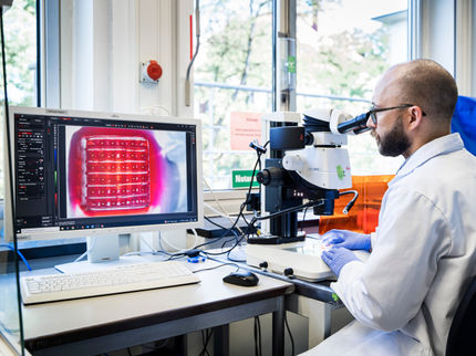
In initial tests, the researchers generated prints with type I collagen inks at concentrations of 3%, 5% and 7% using different nozzles, evaluating structural stability and cell viability. At 3%, the ink continued to spread after printing and during gelation. However, the two higher concentrations enabled freestanding structures to be printed. The team has already successfully printed initial skin tissue models, demonstrating the potential of the type I collagen ink. Real-time monitoring of the human cells’ metabolism also enables the researchers to continuously track the tissue’s nutrient supply. This makes it possible to assess the quality of the printed tissue structures and systematically optimize their architecture.
The new technology marks a major advance. Cost constraints mean that nearly all printing inks currently use animal-derived, highly modified collagens that can replicate human physiology only to a limited extent. In contrast, PhysioINK printing inks will be made from physiological components, ideally human-derived. These are promising conditions for drug research and for printing customized, viable and implantable organs.


Other news from the department science









Most read news







More news from our other portals












Something is happening in the life science industry ...
This is what true pioneering spirit looks like: Plenty of innovative start-ups are bringing fresh ideas, lifeblood and entrepreneurial spirit to change tomorrow's world for the better. Immerse yourself in the world of these young companies and take the opportunity to get in touch with the founders.