World's first high-resolution brain developed by 3D printer
New model can advance research into neurodegenerative diseases
Advertisement


In a joint project between TU Wien and MedUni Vienna, the world's first 3D-printed "brain phantom" has been developed, which is modelled on the structure of brain fibres and can be imaged using a special variant of magnetic resonance imaging (dMRI). As a scientific team led by TU Wien and MedUni Vienna has now shown in a study, these brain models can be used to advance research into neurodegenerative diseases such as Alzheimer's, Parkinson's and multiple sclerosis. The research work was published in the journal "Advanced Materials Technologies".
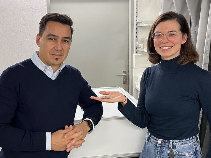
Franziska Chalupa-Gantner with the brain phantom in her hand (right) and Aleksandr Ovsianikov (left).
TU Wien
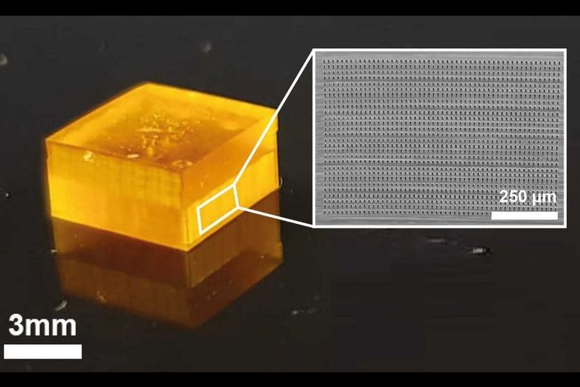
The brain phantom
MedUni Wien

Magnetic resonance imaging (MRI) is a widely used diagnostic imaging technique that is primarily used to examine the brain. MRI can be used to examine the structure and function of the brain without the use of ionising radiation. In a special variant of MRI, diffusion-weighted MRI (dMRI), the direction of the nerve fibres in the brain can also be determined. However, it is very difficult to correctly determine the direction of nerve fibres at the crossing points of nerve fibre bundles, as nerve fibres with different directions overlap there. In order to further improve the process and test analysis and evaluation methods, an international team in collaboration with the TU Wien and the Medical University of Viennadeveloped a so-called "brain phantom", which was produced using a high-resolution 3D printing process.
Tiny cube with microchannels
Researchers from the Medical University of Vienna as MRI experts and TU Wien as 3D printing experts worked closely with colleagues from the University of Zurich and the University Medical Centre Hamburg-Eppendorf. Back in 2017, a two-photon polymerisation printer was developed at TU Wien that enables upscaled printing. In the course of this, work was also carried out on brain phantoms as a use case together with the Medical University of Vienna and the University of Zurich. The resulting patent forms the basis for the brain phantom that has now been developed and is being supervised by TU Wien's Research and Transfer Support team.
Visually, this phantom does not have much to do with a real brain. It is much smaller and has the shape of a cube. Inside it are extremely fine, water-filled microchannels the size of individual cranial nerves. The diameters of these channels are five times thinner than a human hair. In order to imitate the fine network of nerve cells in the brain, the research team led by first authors Michael Woletz (Center for Medical Physics and Biomedical Engineering, MedUni Vienna) and Franziska Chalupa-Gantner (3D Printing and Biofabrication research group, TU Wien) used a rather unusual 3D printing method: two-photon polymerisation. This high-resolution method is primarily used to print microstructures in the nanometre and micrometre range - not for printing three-dimensional structures in the cubic millimetre range. In order to create phantoms of a suitable size for dMRI, the researchers at TU Wien have been working on scaling up the 3D printing process and enabling the printing of larger objects with high-resolution details. Highly scaled 3D printing provides the researchers with very good models that - when viewed under dMRI - make it possible to assign various nerve structures. Michael Woletz compares this approach to improving the diagnostic capabilities of dMRI with the way a mobile phone camera works: "We see the greatest progress in photography with mobile phone cameras not necessarily in new, better lenses, but in the software that improves the captured images. The situation is similar with dMRI: using the newly developed brain phantom, we can adjust the analysis software much more precisely and thus improve the quality of the measured data and reconstruct the neural architecture of the brain more accurately."
Brain phantom trains analysis software
The authentic reproduction of characteristic nerve structures in the brain is therefore important for "training" the dMRI analysis software. The use of 3D printing makes it possible to create diverse and complex designs that can be modified and customised. The brain phantoms thus depict areas in the brain that generate particularly complex signals and are therefore difficult to analyse, such as intersecting nerve pathways. In order to calibrate the analysis software, the brain phantom is therefore examined using dMRI and the measured data is analysed as in a real brain. Thanks to 3D printing, the design of the phantoms is precisely known and the results of the analysis can be checked. TU Wien and MedUni Vienna were able to show that this works as part of the joint research work. The phantoms developed can be used to improve dMRI, which can benefit the planning of operations and research into neurodegenerative diseases such as Alzheimer's, Parkinson's and multiple sclerosis.
Despite the proof of concept, the team still faces challenges. The biggest challenge at the moment is scaling up the method: "The high resolution of two-photon polymerisation makes it possible to print details in the micro- and nanometre range and is therefore very suitable for imaging cranial nerves. At the same time, however, it takes a correspondingly long time to print a cube several cubic centimetres in size using this technique," explains Chalupa-Gantner. "We are therefore not only aiming to develop even more complex designs, but also to further optimise the printing process itself."
Original publication
Michael Woletz, Franziska Chalupa‐Gantner, Benedikt Hager, Alexander Ricke, Siawoosh Mohammadi, Stefan Binder, Stefan Baudis, Aleksandr Ovsianikov, Christian Windischberger, Zoltan Nagy; "Toward Printing the Brain: A Microstructural Ground Truth Phantom for MRI"; Advanced Materials Technologies, Volume 9, 2024-1-7


Other news from the department science