Advances in Cancer Research: New Software Simplifies the Analysis of Complex Tissue Images
Advertisement


Thanks to modern imaging techniques, it is now possible to determine, using just a single tissue sample, which cells are present in the tissue and how they are organized. This breakthrough has significantly simplified the diagnosis and research of various diseases. An interdisciplinary research team from Heinrich Heine University Düsseldorf (HHU), Düsseldorf University Hospital (UKD), and the European molecular biology Laboratory (EMBL) has now developed software designed to assist in the analysis of these results in the future. The researchers have now published their findings in the prestigious journal *Nature Methods*.
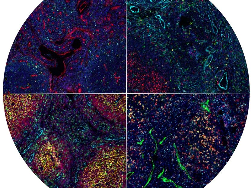
Multiparametric microscopy simultaneously detects numerous proteins in a single tissue sample—in this case, a lymphoma biopsy. In this way, it makes various cell types and tissue structures visible. These can be systematically and reproducibly analyzed using the spatialproteomics software.
Modern microscopy methods, through multiparametric imaging, make it possible to create a kind of map of tissue samples that can be used in the examination of the tissue and, for example, for diagnostic purposes. In doing so, scientists examine various aspects, such as the abundance or spatial distribution of different cell types. This is particularly relevant for cancer research, as cancer cells do not exist in isolation but interact closely with immune cells, blood vessels, and connective tissue cells. The spatial arrangement of these cells can influence how a disease progresses and how effective a treatment is. Unlike traditional examinations, which typically consider only a few features at a time, multiparametric imaging enables the detailed characterization of many thousands to millions of individual cells, thereby simplifying cancer research and diagnosis.
However, analyzing these images is complex: the various cell types must be identified, measured, and counted. Only then can researchers determine which cells exhibit abnormalities, such as spatial clustering. Until now, this process required multiple programs and laboriously customized analysis steps. The new software, spatialproteomics—which researchers led by first author Matthias Meyer-Bender (EMBL) have now presented in the prestigious journal *Nature Methods*—is expected to significantly simplify this process in the future. It enables the various processing steps to be consolidated and supports the analysis of the original microscopy images.
In their paper, Matthias Meyer-Bender, Dr. Peter-Martin Bruch (co-last author, HHU, UKD), and their team demonstrated how the software works using B-cell non-Hodgkin lymphomas as an example. Using the software made it possible to analyze the distribution of different cell types within a biopsy. In this way, spatialproteomics helped distinguish between benign and malignant lymphomas.
“Easily accessible analysis programs are a key prerequisite for the widespread adoption of new imaging technologies. They lower technical barriers to entry, improve the comparability of studies, and make it easier to evaluate complex data in a reproducible manner,” explains first author Meyer-Bender. Bruch adds: “This means that spatialproteomics can not only support lymphoma research but can also be used more broadly to investigate various tumor diseases, inflammatory processes, and other complex tissues.”
Bruch considers the interdisciplinary collaboration between HHU, UKD, and EMBL a particular success. “We were able to combine the clinical and experimental expertise from Düsseldorf with EMBL’s methodological expertise in bioinformatics, data science, and software development. This close collaboration was crucial for creating a tool that is both technically powerful and tailored to specific biological and medical questions,” Bruch says with satisfaction.
Note: This article has been translated using a computer system without human intervention. LUMITOS offers these automatic translations to present a wider range of current news. Since this article has been translated with automatic translation, it is possible that it contains errors in vocabulary, syntax or grammar. The original article in German can be found here.
Original publication


Other news from the department science















These products might interest you




Most read news








More news from our other portals















See the theme worlds for related content
Topic world Diagnostics
Diagnostics is at the heart of modern medicine and forms a crucial interface between research and patient care in the biotech and pharmaceutical industries. It not only enables early detection and monitoring of disease, but also plays a central role in individualized medicine by enabling targeted therapies based on an individual's genetic and molecular signature.

Topic world Diagnostics
Diagnostics is at the heart of modern medicine and forms a crucial interface between research and patient care in the biotech and pharmaceutical industries. It not only enables early detection and monitoring of disease, but also plays a central role in individualized medicine by enabling targeted therapies based on an individual's genetic and molecular signature.
Topic world Fluorescence microscopy
Fluorescence microscopy has revolutionized life sciences, biotechnology and pharmaceuticals. With its ability to visualize specific molecules and structures in cells and tissues through fluorescent markers, it offers unique insights at the molecular and cellular level. With its high sensitivity and resolution, fluorescence microscopy facilitates the understanding of complex biological processes and drives innovation in therapy and diagnostics.

Topic world Fluorescence microscopy
Fluorescence microscopy has revolutionized life sciences, biotechnology and pharmaceuticals. With its ability to visualize specific molecules and structures in cells and tissues through fluorescent markers, it offers unique insights at the molecular and cellular level. With its high sensitivity and resolution, fluorescence microscopy facilitates the understanding of complex biological processes and drives innovation in therapy and diagnostics.