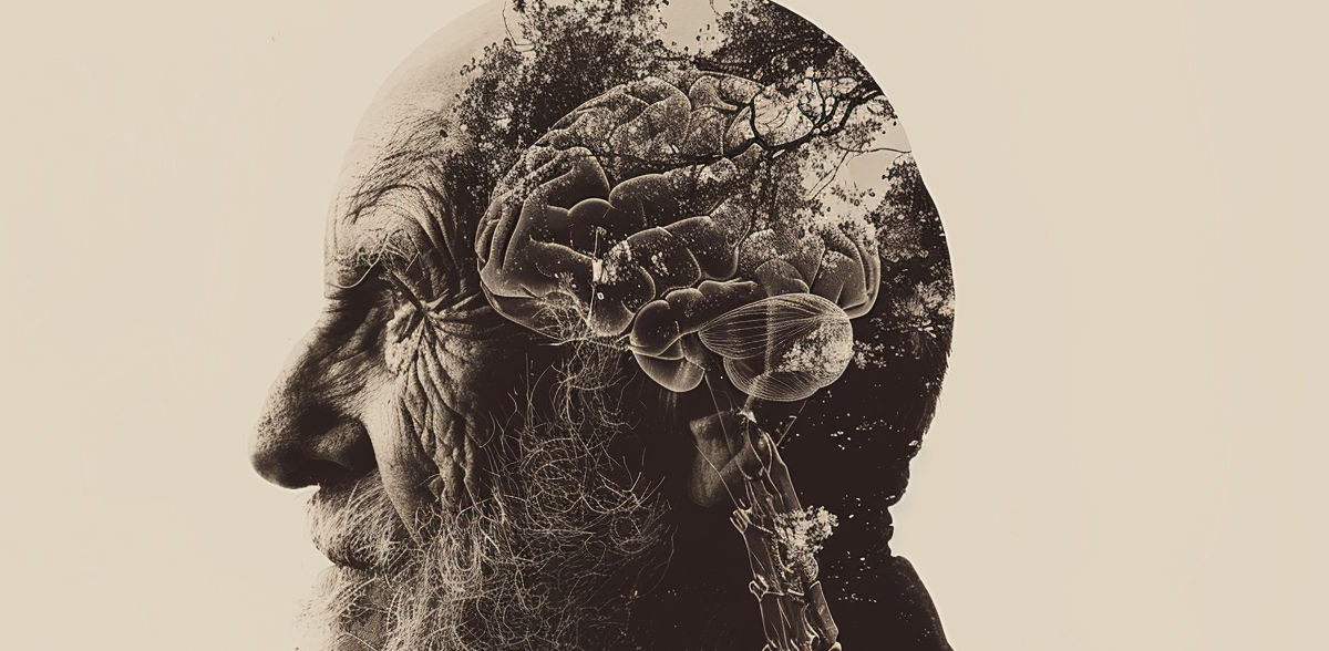
The aging brain: protein mapping furnishes new insights
Researchers at LMU and the SyNergy Cluster of Excellence have analyzed how the proteome of specific brain cells changes as we age
Advertisement


For the neurons in the brain to work smoothly and be able to process information, the central nervous system needs a strictly regulated environment. This is maintained by the blood-brain barrier, whereby specialized brain endothelial cells lining the inner walls of blood vessels regulate the exchange of molecules between the circulatory and nervous systems. Earlier studies have shown that various functions that are dependent on these cells, such as the integrity of the blood-brain barrier or the regulation of blood supply to the brain, decline over the course of a person’s life. This dysregulation leads to a dysfunction of the brain vasculature and is therefore a major contributor to medical conditions such as strokes and dementia.
However, the molecular changes that underlie this loss of function have remained largely obscure. To improve our mechanistic understanding, researchers carry out molecular profiling studies to investigate the different components of brain endothelial cells and collect their findings in large databases. “The transcriptome – that is to say, the RNA contained in endothelial cells – has since been quite comprehensively mapped,” says LMU professor Martin Dichgans, Director of the Institute for Stroke and Dementia Research at University of Munich Hospital and Principal Investigator at the SyNergy Cluster of Excellence. “What has been lacking is corresponding data on the complete set of proteins in the cells, the proteome.” A study recently published in the journal Nature Aging, which had major contributions by researchers from LMU and SyNergy, has now closed this knowledge gap.
Dysregulated metabolism
For the study, the team developed a protocol for enriching brain endothelial cells in mice, which makes it possible to resolve age-related changes in protein composition. Using an unsupervised (computer-aided) cluster analysis, the scientists then related these protein dynamics to biological functions. “Our results show a dysregulation of key molecules involved in the uptake of substances into cells, in receptor recycling, and in the degradation of molecules within specific cellular compartments called lysosomes,” says Dichgans.
One of the most striking changes concerned a decrease in proteins involved in vesicle-mediated transport. In addition, the study provides evidence that deficiency of apolipoprotein E, a protein involved in lipid metabolism results in a signature of accelerated endothelial aging. “The results complement and expand findings from studies on the RNA sequencing of brain endothelial cells during aging,” summarizes Dichgans. “Our proteomic approach captures processes that are not detected at the RNA level.” Overall, the study offers a framework for understanding important endothelial signaling pathways during aging and serves as a data basis for future analyses of brain endothelial function. The researchers are making their data on age-related protein abundance of the mouse endothelium available in a publicly accessible database for further use.
Original publication
Katalin Todorov-Völgyi, Judit González-Gallego, Stephan A. Müller, Nathalie Beaufort, Rainer Malik, Martina Schifferer, Mihail Ivilinov Todorov, Dennis Crusius, Sophie Robinson, Andree Schmidt, Jakob Körbelin, Florence Bareyre, Ali Ertürk, Christian Haass, Mikael Simons, Dominik Paquet, Stefan F. Lichtenthaler, Martin Dichgans; "Proteomics of mouse brain endothelium uncovers dysregulation of vesicular transport pathways during aging"; Nature Aging, 2024-3-22


Other news from the department science


