Spiky insight: How red blood cells deform
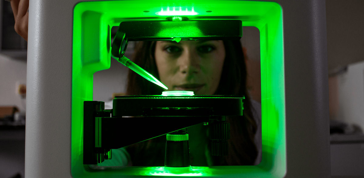
Holotomographic microscopy
Advertisement


Empa researchers have observed living red blood cells transforming into spiky "echinocytes" in real time when treated with high concentrations of ibuprofen using holotomographic microscopy and displayed them in 3D renderings.
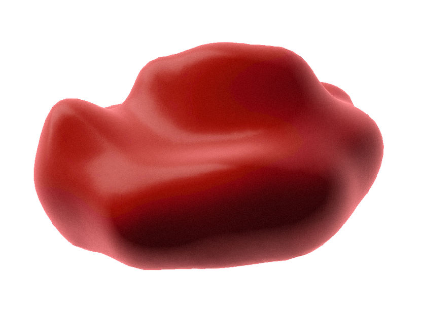
After contact with ibuprofen, the blood cells form bumps and spikes and become echinocytes.
Empa
Blood is indeed "a juice of rarest quality." What the poet and natural scientist Goethe already suspected can now actually be visualized using innovative imaging techniques. One of these special features is the cell that occurs most frequently in the bloodstream: the erythrocyte. Trillions of these red blood cells make their way through the human body every minute. The fact that they do not always take on a round shape enables them to squeeze through the narrowest blood vessels to supply the most remote corners of our body with oxygen.
However, some changes in the shape of erythrocytes are also typical of special changes in the environment: so-called echinocytes with spiny extensions similar to a sea urchin occur, for instance, in the case of burns, liver damage, or after contact with certain drugs. Empa researchers have now observed the transformation of red blood cells into echinocytes using digital holotomographic microscopy.
Talia Bergaglio and Peter Nirmalraj from Empa's Transport at Nanoscale Interfaces laboratory in Dübendorf provoked the deformation of living red blood cells by adding the drug ibuprofen. They were able to show the transformation of roundish donuts into echinocytes in real time thanks to holotomographic microscopy. This innovative technique works similarly to computed tomography (CT), where imaging takes place via laser technology instead of X-rays. Digital holotomographic microscopy is therefore particularly suitable for biological samples such as blood cells, as it enables high-resolution, non-contact and marker-free images to be taken, which can then be reconstructed as three-dimensional representations.
Red blood cells are ideal model cells for this purpose, as they can be easily identified even in whole blood and given their morphology is sensitive to chemical and physical environment, during the course of their existence; they are ultimately (almost) empty membrane shells. "Therefore, the interactions of a variety of drug molecules with the cell membrane can be studied particularly well on red blood cells using our bio-imaging technique," says Empa researcher Nirmalraj.


Other news from the department science


