Unprecedented dynamic-range resolution useful for examining impact of environmental pollutants on cells and tissues
Full-body synchrotron imaging of roundworms reveals nanoparticle accumulation
Advertisement


Scientists working at DESY’s PETRA III light source have been able to generate the first synchrotron-based X-ray map of the whole body of an organism at such a high resolution that contaminants could be detected at the individual cell level. The research team was able to identify cerium nanoparticles in the digestive system of the roundworm Caenorhabditis elegans while also determining that the nanoparticles showed relatively low toxicity. The results from PETRA III were corroborated by fluorescent microscopy, and the method behind the result could prove valuable also for assessment of other environmental pollutants. The results were published in the journal Environmental Science and Technology and involved scientists from the Norwegian University of Life Sciences and the University of Antwerp from the Netherlands.
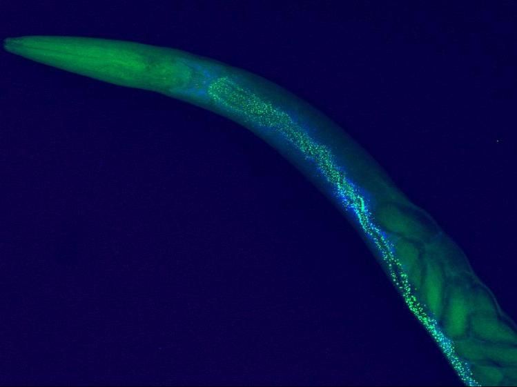
Cerium nanoparticles are illuminated along the head of a C.elegans roundworm in data taken at PETRA III. The worm was exposed to much higher concentrations of cerium than occur in nature to clearly see the uptake of the nanoparticles into the organism. The data set from PETRA III's P06 beamline can also be enlarged to a scale of 300 nanometres.
Norwegian University of Life Sciences (NMBU)
Nanoparticles exist in various forms in the environment. They are defined as any matter particles that measure below the threshold of a millionth of a metre in width. This research investigated nanoparticles of the rare earth metal cerium, which are among the most manufactured nanomaterials due to their unique properties and application in automobile catalytic converters, fuel cells, and materials that block ultraviolet light. The cerium nanoparticles have thus been detected in the wider environment around industrial sites. The effects of release of such nanomaterials in the environment is uncertain, with previous studies showing inconsistent results on the particles’ potential toxicity.
Ole Christian Lind from the Norwegian University of Life Sciences (NMBU) has been studying a variety of environmental contaminants at synchrotron sources such as PETRA III for the past two decades. Normally, such synchrotron studies are performed on preserved, usually dried, specimens. To reach the goal of performing a full imaging study on an undried specimen, Lind used PETRA III’s P06 beamline, headed by DESY scientist Gerald Falkenberg. As the sample, the scientists used the roundworm Caenorhabditis elegans. “We used C. elegans because it is perhaps the most studied eukaryote model organism on the planet,” says Lisa Rossbach, a postdoc at NMBU who is one of the study’s primary authors.
The scientists exposed the worms to a high concentration of cerium nanoparticles, much higher than those found in the environment. “Using X-ray fluorescence spectroscopy, we managed to piece together an extremely high-resolution picture of roundworms that were preserved in a pristine condition,” says Falkenberg. “We accomplished this by using an extremely narrow X-ray beam to excite the electrons in clusters of the nanoparticles and the worm’s tissues, allowing a full internal picture of the organism and the cerium distribution within it.” The data were so detailed that they allow scientists to view the entire organism while also zooming in on individual parts of cells, allowing the scientists to localise clusters of nanoparticles within.
Lind says: “This is a breakthrough experiment. The ability to map uptake of contaminants at 300-nanometre resolution for full-body X-ray imaging of an intact non-dehydrated organism enables us to address the consequences of pollutants in a totally new way. This is next-level toxicology, where we can in detail show that the presence of a contaminant in a tissue or a cell is directly connected to the effect that it generates.”
The pictures revealed that high concentrations of cerium nanoparticles did not cause acute toxic effects and cleared quickly from the body. “However, cerium did accumulate in the mouthparts and caused deformities to the worms’ food processing apparatus”, says Rossbach. As part of this study, Rossbach also investigated the same tissue malformations using in vivo fluorescence microscopy, corroborating the effect of the nanoparticles by examining levels of oxidative stress in the tissues.
The results are also if great interest because cerium is often used as an analogue for radioactive pollutants, which themselves often present themselves as nanoparticles. “In addition to being an interesting result in its own, this paves the way for far more detailed studies on the effects of various kinds of pollution on organisms.” Future experiments could also focus on nanoplastics, which are appearing in even some of the most remote areas of the environment.
Original publication
Synchrotron XRF Analysis Identifies Cerium Accumulation Colocalized with Pharyngeal Deformities in CeO2 NP-Exposed Caenorhabditis elegans; Lisa Magdalena Rossbach, Dag Anders Brede, Gert Nuyts, Simone Cagno, Ragni Maria Skjervold Olsson, Deborah Helen Oughton, Gerald Falkenberg, Koen Janssens, and Ole Christian Lind; Environmental Science and Technology, 2022


Other news from the department science


Most read news


More news from our other portals


See the theme worlds for related content
Topic World Spectroscopy
Investigation with spectroscopy gives us unique insights into the composition and structure of materials. From UV-Vis spectroscopy to infrared and Raman spectroscopy to fluorescence and atomic absorption spectroscopy, spectroscopy offers us a wide range of analytical techniques to precisely characterize substances. Immerse yourself in the fascinating world of spectroscopy!