Study reveals effects of chemoradiation in brains of glioblastoma patients
Advertisement


A study from Massachusetts General Hospital (MGH) Cancer Center researchers - the first to examine the effects of combined radiation and chemotherapy on the healthy brain tissue of glioblastoma patients - reveals not only specific structural changes within patients' brains but also that the effect of cancer therapy on the normal brain appears to be progressive and continues even after radiation therapy has ceased. The report appears in Neurology.
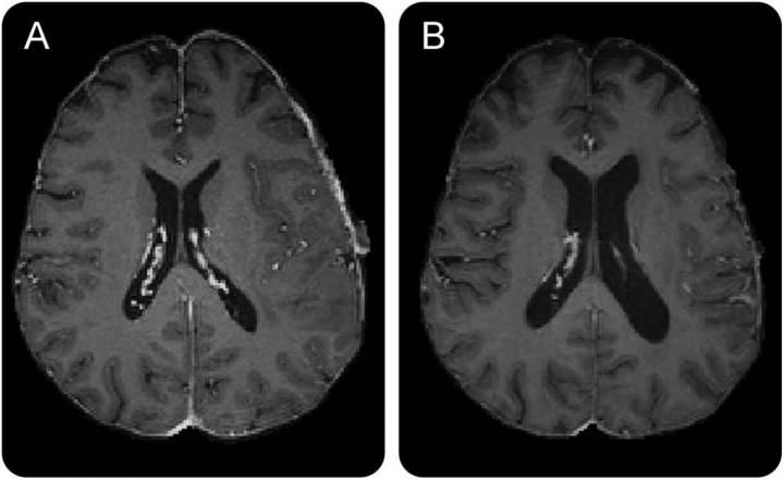
Neurology, ©2015 American Academy of Neurology, Used by permission.
"It is well known that whole brain radiation can have adverse, neurotoxic effects and causes loss of brain volume in some individuals," says Jorg Dietrich, MD, PhD, of the Pappas Center for Neuro-Oncology at MGH. "This is the first prospective and longitudinal study to characterize structural brain changes resulting from standard radiation and chemotherapy in patients with malignant brain tumors. Further studies with neuropsychological evaluation will be needed to characterize the functional consequences of these structural changes."
The study enrolled 14 glioblastoma patients who were scheduled to receive chemotherapy and radiation after surgical tumor removal. Before and during the 35-week standard treatment protocol MR images were taken. In the 8 participants for whom an adequate number of imaging studies were completed, whole brain volume decreased significantly throughout the study period. The reduced volume was apparent within a few weeks after initiation of treatment and was primarily seen in grey matter. The size of the brain's ventricles - cerebrospinal fluid-filled spaces deep within the brain - became progressively larger during the course of treatment, and changes were also seen within the subventricular zone, one of two structures in which new brain cells are generated in adults.
"We were surprised to see that these changes - reduced grey matter volume and ventricular enlargement - occurred after just a few weeks of treatment and continued to progress even after radiation therapy was completed," says Dietrich. "While this was a small study, these changes affected every patient at least to some degree. Now we need to investigate whether these structural changes correlate with reduced cognitive function and whether neuroprotective strategies might be able to stop the progression of brain volume loss. Establishing novel imaging biomarkers of treatment-associated neurotoxicity - such as ventricular enlargement, which can be tracked with any MR scanner - will be a critical step towards developing more selective therapies that are targeted to the tumor and spare normal brain tissue."


Other news from the department science


