Medically relevant nanoparticles move faster in cells than expected
Finding has large implications for applications in nanomedicine such as targeted drug delivery and intracellular studies
Advertisement


An experiment led by a team of DESY and Universität Hamburg scientists has discovered that gold nanoparticles can move through liquid biological matter faster than expected when coated by the polymer polyethylene glycol (PEG). The team’s data, which were acquired using X-ray photon correlation spectroscopy, reveal both the structure and the dynamics of the nanoparticles in various biological fluids with high temporal resolution. The research has been published in the journal Aggregate.
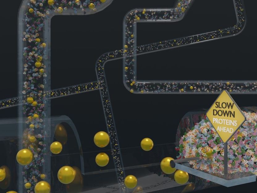
To illustrate the type of environments in which gold nanoparticles can be used, the nanoparticles approach densely packed regions of proteins. The researchers behind this study found that the nanoparticles, when coated with the polymer PEG, move quickly through such densely packed environments.
Ferdinand Otto, UHH
In the ever-expanding field of nanomedicine, gold nanoparticles have become a promising tool. The nanoparticles are tiny gold crystals that are surrounded by organic molecules, which are called ligands. The coating of gold nanoparticles with such ligands can be precisely controlled. Additionally, gold is non-toxic, and gold nanoparticles have useful physical properties like strong absorption and scattering of visible light and X-rays. This makes gold nanoparticles very useful for medical applications. They found widespread use in the COVID-19 pandemic as the key ingredient in self-test cartridges used worldwide. They also show high potential for targeted drug delivery, as contrast agents for computed tomography, and for hyperthermal therapy approaches, among others. For instance, as a potential cancer treatment, they can be injected into a tumour site and then be superheated with a laser in order to kill the cancer cells from within.
Many approved nanoparticles for medical applications, including the lipid nanoparticles used for most COVID vaccines, are coated with PEG to improve their stability, solubility, and distribution in the body. Understanding how particle dynamics in biological fluids are affected by this polymer is therefore of utmost importance. Owing to their tiny size, the nanoparticles can even enter the target cells, usually once they are surrounded by a clump of proteins, which are present in high concentrations in blood and at even higher densities in the interior of cells. The clump of proteins not only changes the properties of the nanoparticles so that they might not reach their target anymore, but it also can trigger an undesirable immune response. PEG coatings can strongly reduce this unwanted protein adsorption, and scientists often talk about a “stealth” effect provided by the PEG coating.

In their study, the team used gold nanoparticles with tailored PEG-coatings and studied their dynamics in the presence of a varying protein concentrations. This way, they were able to mimic the different environments nanoparticles encounter in the body: from the bloodstream to the interior of cells with extremely high protein concentrations.
“The more molecules you put into the fluid, the more molecularly dense it is, the closer you get to what’s called protein crowding, which is what’s happening in the body,” says Felix Lehmkühler, a scientist at DESY who co-led the research. Lehmkühler likens protein crowding to a big party in a small room: the party attendees are all standing close to each other, and someone– in this case, the gold nanoparticle – trying to get through has to continually change direction or is being stopped by some of the partygoers on the way.
“Past experiments have consistently shown this protein adsorption process to be a common effect. We wanted to push our experiment to the extremes of what was possible, to a high protein concentration similar to real life, and test the effects of a PEG shell” says Florian Schulz, a postdoc at Universität Hamburg who co-led the experiment with Lehmkühler. “We would’ve expected to see that the nanoparticles slow down once they enter protein-rich environments.”
However, the team found something much different in their results. “When we come into this protein crowding situation, we find that the particle is moving faster than expected,” says Lehmkühler. It seems as if the nanoparticle speedily cut through the party in the previous analogy. The team used X-ray photon correlation spectroscopy to monitor the diffusion of the nanoparticles. They observed no indications of protein adsorption to the nanoparticles as they move around, thus confirming the beneficial “stealth” effect of the PEG-coatings.
In very crowded protein environments, similar to the situation in cells, the nanoparticles diffuse much faster than the viscosity predicted by theory would allow. Even more fascinatingly, the team found that the viscosities of the protein-rich biological solutions are exactly as expected when measured with classical methods. As a result, the viscosity local to the nanoparticle strongly differs from the viscosity of the biological material as a whole – meaning that the nanoparticle has a strong effect on its surrounding that enables this speedy motion. This is an extremely important finding for the understanding of nanoparticle dynamics in biological systems, which is key for their safe and successful application.
“The nanoscale viscosity changes were a surprise,” says Schulz. “That is the first time this phenomenon has been observed. We had to check and check and double check this to make sure this effect was real. This is a really important finding, because it tells us that these nanoparticles diffuse so quickly because they strongly affect their local environment.”
“This study underlines how important these thorough measurements of structure and dynamics are for our understanding of complex systems”, says Ferdinand Otto, the first author of the publication, “and X-ray photon correlation spectroscopy is an excellent method that is just at the beginning of being explored in this context.”
The experiment was performed at the European Synchrotron Radiation Facility (ESRF) in Grenoble, France. This type of research, however, will benefit highly from the planned PETRA IV light source at DESY. The technique used in this research, X-ray photon correlation spectroscopy, will be the technique that gains the most from the facility upgrade, as the brilliance of PETRA IV will enable an extension of the accessible timescale to nanoseconds, or a billionth of a second.
Original publication


Other news from the department science


Most read news


More news from our other portals


See the theme worlds for related content
Topic World Spectroscopy
Investigation with spectroscopy gives us unique insights into the composition and structure of materials. From UV-Vis spectroscopy to infrared and Raman spectroscopy to fluorescence and atomic absorption spectroscopy, spectroscopy offers us a wide range of analytical techniques to precisely characterize substances. Immerse yourself in the fascinating world of spectroscopy!