Accurate data on dynamic molecular aggregates in cells for the first time
Using a newly developed microscopy method, researchers have for the first time been able to quantify small, dynamic molecular aggregates in living cells that play an important role in signal processing
Advertisement


In cells, many vital processes take place in membrane-free molecular aggregates. Such aggregates ensure that the molecules involved are present in the right concentration and proximity to each other. Researchers from the Cluster of Excellence CIBSS at the University of Freiburg and the University of Cambridge/UK have now been able to observe and analyze the formation of these so-called condensates in living cells for the first time. In Nature Communications, they describe that not only physical principles, but also active control mechanisms regulate their growth. The experimental protocols and analysis programs are now available free of charge, making research on small aggregates possible even for less specialized laboratories.
If the molecules inside a cell were randomly distributed, it would not be viable. Only by dividing them into specialized compartments can many biochemical processes take place in a coordinated manner. In part, these compartments are separated from each other by membranes, but a separation also functions without an outer shell. Such membrane-free molecular aggregates, also called condensates, fulfill important biological functions because their size and number are particularly flexible. They are thought to form through the physical process known as liquid-liquid phase separation.
"These condensates are an important control mechanism in cells because they can accelerate or slow down biochemical processes as needed," explains Prof. Dr. Thorsten Hugel. He is a member of the Cluster of Excellence CIBSS - Centre for Integrative Biological Signalling Studies at the University of Freiburg and co-led the current study with Prof. Dr. Aleks Reinhardt from the University of Cambridge.
Smaller condensates are more difficult to study
How condensates help the cell process biological signals and environmental stimuli is still far too little studied, Hugel says. "People usually focus on large and static condensates in research because they're easier to study. But these large condensates are usually just the end stage of a long process. Much more interesting are small condensates that grow and decay dynamically," he explains. The problem: They consist of relatively few molecules and are too small and fast even for high-resolution microscopy methods to study them in living cells.
New method circumvents technical limitations
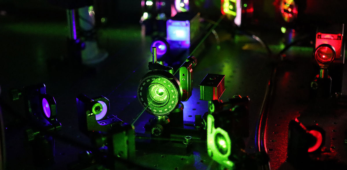
In the current study, the researchers from Freiburg and Cambridge have now described a way to circumvent these technical limitations. To do so, they used conventional high-resolution fluorescence microscopy with an oblique laser, so-called HILO microscopy, and combined it with a special experimental approach and AI-based analysis methods.
Growth cannot be explained by physical processes alone
The researchers compared the measurements thus made in living cells with theoretical assumptions about the formation of condensates. "The results surprised us," says Reinhardt, who is a researcher in the Department of Chemistry at the University of Cambridge. "For the condensates we investigated in this study, the initial growth still follows physical models. We would expect the same for such processes. But then, above a certain size, this growth suddenly stops."
Growth of NELF aggregates is regulated by stress signals
In the current study, the researchers examined aggregates of the protein NELF. These protein aggregates form when a cell is stressed by heat, for example, or when aggregates of other proteins form, as in dementia and other neurogenerative diseases. "By forming condensates in the nucleus, NELF inhibits the expression of genes," said co-author Dr. Ritwick Sawakar, summarizing the protein's natural function. "This inhibition is important for the cell to survive stress." Sawakar also worked at CIBSS and currently conducts research at the MRC Toxicology Unit at the University of Cambridge.
The scientists* have now observed that many small NELF condensates are present in cells even in the absence of stress. "Outside a cell, we would expect the condensates to continue to grow once they reach a critical size. In living cells, however, they seem to do so only when the cell is under stress," Reinhardt describes the result. The researchers conclude that NELF condensates are actively kept small until stress signals release the growth again.
Protein aggregates are probably an important mechanism of signal processing
According to the scientists*, this process, which seems complicated at first glance, is probably essential for the processing of stress signals: "Larger condensates can thus form particularly quickly when required and small ones can dissolve very quickly," Hugel explains. "This allows the cell to respond to stress in a timely manner."
Protein aggregates are thought to have many different and fundamental functions for signal processing in cells. The new microscopy method now makes it possible to understand these functions comprehensively. This also makes it possible to explore the role of protein aggregates in diseases such as dementia, Alzheimer's and Huntington's. In the long term, a better understanding of this could also help in the diagnosis or development of therapies for such diseases.
Note: This article has been translated using a computer system without human intervention. LUMITOS offers these automatic translations to present a wider range of current news. Since this article has been translated with automatic translation, it is possible that it contains errors in vocabulary, syntax or grammar. The original article in German can be found here.
Original publication
Chenyang Lan, Juhyeong Kim, Svenja Ulferts, Fernando Aprile-Garcia, Sophie Weyrauch, Abhinaya Anandamurugan, Robert Grosse, Ritwick Sawarkar, Aleks Reinhardt, Thorsten Hugel; "Quantitative real-time in-cell imaging reveals heterogeneous clusters of proteins prior to condensation."; Nature Communications (2023).


Other news from the department science


Most read news


More news from our other portals


See the theme worlds for related content
Topic world Fluorescence microscopy
Fluorescence microscopy has revolutionized life sciences, biotechnology and pharmaceuticals. With its ability to visualize specific molecules and structures in cells and tissues through fluorescent markers, it offers unique insights at the molecular and cellular level. With its high sensitivity and resolution, fluorescence microscopy facilitates the understanding of complex biological processes and drives innovation in therapy and diagnostics.