Synapses in 3D
Scientists develop new method to map brain structures
Advertisement


Our brain consists of countless nerve cells that transmit signals from one cell to the next. The connections between these cells, the synapses, provide a key to understanding how our memory works. An American research team in collaboration with Rainer Heintzmann from the Leibniz Institute of Photonic Technology (Leibniz IPHT) and the Friedrich Schiller University Jena has now succeeded in identifying these switching points in millimeter-sized tissue with a light microscope on the basis of their structure.
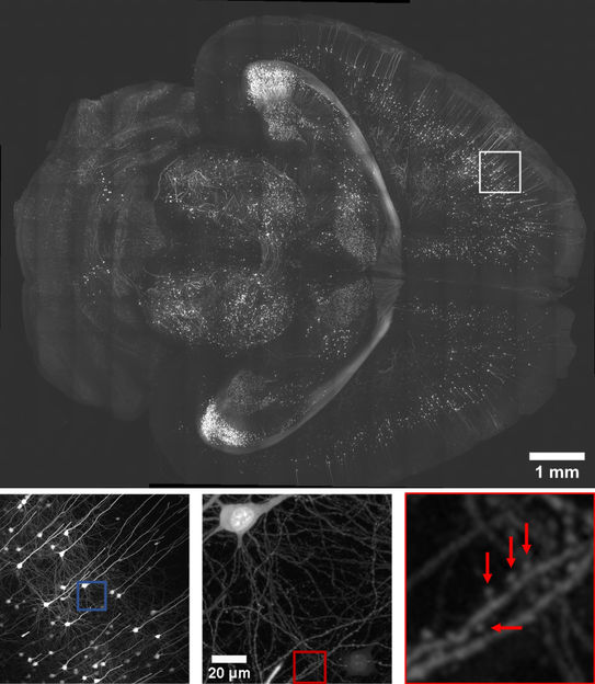
An x–y view of a section 2.5 mm from the top surface of a Thy1-eGFP PEGASOS-cleared brain. Close-up of the box with a rendered neuron. Insets provide magnified views of synaptic spines.
Reto Fiolka
To make the synapses visible, the research team at Southwestern University Texas, led by Reto Fiolka and Kevin Dean, developed a special microscope. The scientists illuminate a tissue sample approximately one millimeter in size from the side with wedge-shaped focused light. While the focus of this light wedge shifts, image data is recorded. This enables the researchers to use machine learning to identify and visualize three-dimensional tissue structures within the cells in high resolution and true to scale. Depending on the optical configuration, the microscope provides up to 260 nm of axial resolution, a three to tenfold improvement over confocal and other reported cleared-tissue light-sheet microscopes. The research team imaged millimeter-scale cleared tissues with subcellular three-dimensional resolution, which enabled automated detection of multicellular tissue architectures, individual cells, synaptic spines and rare cell–cell interactions.
"This work is groundbreaking. The recognition of synapses in millimeter-sized tissue using the light microscope only on the basis of their structure has long been a dream of scientists," said Rainer Heintzmann from Leibniz IPHT. He calculated the expected light distribution and thus the quality of the wedge focus. "The calculations are important for the optical design of the instrument," explains Rainer Heintzmann. "They take into account the unwanted influence that the non-ideal refractive index of the embedding medium has on the quality of the focus. "
The research team believes their method will expedite human cell atlas efforts, providing much- needed insight into how tissue function manifests in health and disease, from the heterogenous cellular populations that compose it. Scientists around the world are working on three-dimensional imaging and characterization of all cells in the human body. The Human Cell Atlas is intended to create comprehensive reference maps of all human cells to contribute to a better understanding of how fundamental processes in our organism take place and how they change when we become ill, thus helping to enable better diagnosis and treatment.
Original publication


Other news from the department science


