New optical method promises faster, more accurate diagnosis of breast cancer
Researchers report on data-based method to replace manual inspection of tissue
Advertisement


A new optical method for more quickly and accurately determining whether breast tissue lesions are cancerous is described by University of Illinois researchers.

Figure 3 from a new article in the Journal of Biomedical Optics compares stained bright-field microscopy (top row) and SLIM (bottom row) images in their respective abilities to show malignant and benign. The images were obtained from adjacent sections. Color bars are in radians.
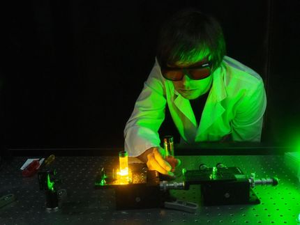
Hassaan Majeed, Univ. of Illinois, et al./SPIE
University of Illinois at Urbana-Champaign and Chicago researchers Hassaan Majeed, Mikhail Kandel, Kevin Han, Zelun Luo, Virgilia Macias, Krishnarao Tangella, Andre Balla, and Gabriel Popescu report on a quantitative method for diagnosing breast cancer using spatial light interference microscopy (SLIM).
Because this method is based on quantitative data rather than a subjective assessment, the researchers expect that these preliminary results show the potential of their technique to become the basis for an automated image analysis system that would provide a fast and accurate diagnostic method.
When an abnormality in the breast is discovered, standard practice is for the physician to take a tissue biopsy, which is then stained to provide enough contrast for a pathologist to study key features of the tissue under a microscope. The tissue analysis is done manually. Due to variations such as staining intensity and the illumination used, the process does not lend itself to automation.
Manual inspections, however, are subject to investigator bias, and the process is time-consuming. This can, in some cases, result in late diagnosis - a critical shortcoming given that early diagnosis significantly improves chances of survival.
Using the breast tissue biopsies of 400 different patients, researchers selected two parallel, adjacent sections from each biopsy. One was stained and the other left unstained.
The unstained samples were analyzed using a SLIM module attached to a commercial phase contrast microscope to generate interferograms.
Four interferograms were used to produce one quantitative image showing areas with different refractive properties in different colors. The boundary between tumors and the cells around them were clearly delineated, making it possible to assess whether the tumors were malignant or benign.
Original publication


Other news from the department science


