Protein critical to gut lining repair
Advertisement


Scientists at Washington University School of Medicine in St. Louis have identified a protein essential to repairing the intestine's inner lining. That lining is among the body's busiest highways, trod not only by the food we ingest but also by trillions of microorganisms that aid digestion. Because the intestine plays key roles in absorbing nutrients and containing the microbes, any damage must be fixed promptly.
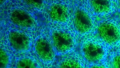
Scientists have identified a protein that is critical to repairing glands in the intestine’s inner lining. The glands appear as dark green spots above and are rebuilt every two to four weeks as the inner lining of the gut is continually renewed.
Hiroyuki Miyoshi
The researchers report in Science Express that a protein called Wnt5a is essential for reconstructing glands in the intestinal lining. The glands, called crypts of Lieberkühn, contain stem cells that continually pump out other cells that renew the gut lining, which is replaced every two to four weeks. The crypts look like dimples in the gut lining and are vulnerable to damage and loss from infection and inflammation.
"For example, inflammatory bowel disease can destroy huge stretches of the lining, including the crypts," says senior author Thaddeus Stappenbeck, MD, PhD, associate professor of pathology and immunology. "If crypts can't be repaired as the lining is rebuilt, their absence would place substantial stresses on crypts in healthy portions of the gut. So it's important to better understand how the crypts are replaced."
In the new study, Stappenbeck and his colleagues showed that when crypts are lost to injury in mice, the nearest surviving crypts expand into the damaged area and create an array of channels. These wound channels, which contain rapidly dividing stem cells, eventually subdivide into new crypts.
Stappenbeck found that cells that line the outer wall of the gut migrate to sites of damage within the inner lining to provide Wnt5a, a signaling molecule that stops stem cells from dividing. This shutdown triggers the formation of dimples in the wound channels that become new crypts.
Because mice that lack the Wnt5a gene aren't viable, co-author Terry Yamaguchi, PhD, of the National Cancer Institute, bred mice in which scientists could selectively turn off the gene after the mice became adults. When the gut lining was injured and Wnt5a was disabled, the wound channels formed but failed to divide into crypts.
To further confirm the link between the gene and crypt repair, Hiroyuki Miyoshi, PhD, a postdoctoral fellow in Stappenbeck's laboratory, devised a robust method for growing gut stem cells in test tubes. When he applied Wnt5a to the stem cells, they stopped dividing, proving that the protein was the critical ingredient for initiating crypt formation.
The scientists also showed that Wnt5a activated a signaling pathway in the stem cells that is known to stop cell proliferation. Stappenbeck, who also is an associate professor of developmental biology, is now planning additional studies of Wnt5a, including investigations of whether the protein plays similar roles elsewhere in the body.
Original publication
Miyoshi H, Ajima R, Luo C T-Y, Yamaguchi TP, Stappenbeck TS; "Wnt5a potentiates TGF-beta signaling to promote colonic crypt regeneration after tissue injury."; Science Express, 2012.


Other news from the department science


