IBN's 'Fish and Chips' May Help Accelerate Drug Discovery
Advertisement


A cheaper, faster and more efficient platform for preclinical drug discovery applications has been invented by scientists at the Institute of Bioengineering and Nanotechnology (IBN). Called 'Fish and Chips', the novel multi-channel microfluidic perfusion platform can grow and monitor the development of various tissues and organs inside zebrafish embryos for drug toxicity testing. This research was published recently in Lab on a Chip.
Current drug studies on zebrafish embryos are performed on traditional microtiter plates, which do not allow perfusion or the replenishment of growth media and drugs, and cannot facilitate live imaging since the embryos are not fixed in one position due to the size of the well. The conventional way of visualizing tissues and organs in embryos is a laborious process, which includes first mounting the embryos in a viscous medium such as gel, and then manually orienting the embryos using fine needles. The embryos also need to be anesthetized to restrict their motion and a drop of saline needs to be continuously applied to prevent the embryos from drying. These additional precautions could further complicate the drug testing results.
The IBN 'Fish and Chips' has been designed for dynamic long-term culturing and live imaging of the zebrafish embryos. The microfluidic platform comprises three parts: 1) a row of eight fish tanks, in which the embryos are placed and covered with an oxygen permeable membrane, 2) a fluidic concentration gradient generator to dispense the growth medium and drugs, and 3) eight output channels for the removal of the waste products. The novelty of the 'Fish and Chips' lies in its unique diagonal flow architecture, which allows the embryos to be continually submerged in a uniform and consistent flow of growth medium and drugs, and the attached gradient generator, which can dispense different concentrations of drugs to eight different embryos at the same time for dose-dependent drug studies.
Other key design elements include customizing the dimensions of the fish tank, which is over 200 times smaller than the individual well of a microtiter plate, to fit the embryos exactly in the tank and restrict its movement for live imaging. This is crucial to monitor the growth and development of the various tissues and organs in the embryos. The microfluidic structure was also fabricated using silicon and glass, which provide greater accuracy and reproducibility for commercial application, in comparison with polydimethylsiloxane (PDMS) polymeric-based chips.
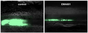
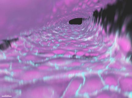
Using high-resolution bright-field and fluorescence imaging, the researchers were able to observe the development of various organs such as the eyes, ears, melanophores, brain, yolk sac, trunk and chorion, as well as heartbeats in the zebrafish embryos. The researchers also conducted drug toxicity testing on the 'Fish and Chips' with valproic acid (VPA), a drug which causes birth defects if consumed by women during their pregnancy. VPA caused abnormality in the development of the eyes and tail in the embryos. The findings clearly established the proof-of-concept that IBN's 'Fish and Chips' could be used as an organ-level drug screening model.
Professor Hanry Yu, IBN Group Leader, who led the research efforts at IBN, said, "Toxicity is a major cause of drug failures in clinical trials and our novel 'Fish and Chips' device can be used as the first step in drug screening during the preclinical phase to complement existing animal models and improve toxicity testing. The design of our platform can also be modified to accommodate more zebrafish embryos, as well as the embryos of other animal models. Our next step will involve investigating cardiotoxicity and hepatoxicity on the chip."


Other news from the department research and development