The “Great Unified Microscope” can see both micro and nanoscale structures
Researchers unify two conventional techniques that so far have been used to make either micro- or nanoscale observations
Advertisement


Researchers Kohki Horie, Keiichiro Toda, Takuma Nakamura, and Takuro Ideguchi of the University of Tokyo have built a microscope that can detect a signal over an intensity range fourteen times wider than conventional microscopes. Moreover, the observations are made label-free, that is, without the use of additional dyes. This means the method is gentle on cells and adequate for long-term observations, holding potential for testing and quality control applications in the pharmaceutical and biotechnology industries. The findings are published in the journal Nature Communications.

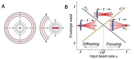
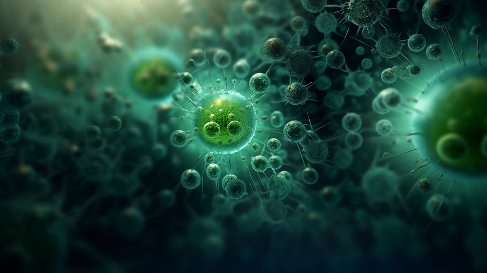
Conceptual illustration of the bidirectional quantitative scattering microscope, which detects both forward and backward scattered light from cells. This dual detection enables visualization of structures ranging from whole-cell morphology to nanoscale particles.
Horie et al 2025
Microscopes have played a pivotal role in the development of science since the 16th century. However, progress has required not only more sensitive and accurate equipment and analysis, but also more specialized ones. Therefore, modern, cutting-edge techniques have had to straddle tradeoffs. Quantitative phase microscopy (QPM) leverages forward-scattered light and can detect structures at the microscale (in this study, over 100 nanometers), but not smaller. Consequently, this technique has been primarily used to take static pictures of relatively complex cell structures. Interferometric scattering (iSCAT) microscopy, on the other hand, exploits back-scattered light and can detect structures as small as single proteins. As such, it can be used to “track” single particles, allowing insight into dynamical changes within the cell, but it cannot provide the comprehensive view that QPM can.
“I would like to understand dynamic processes inside living cells using non-invasive methods,” says Horie, one of the first authors.
Thus, the research team set out to investigate whether measuring both directions of light simultaneously could overcome the tradeoff and reveal a wide range of sizes and motions from the same image. To test the idea and confirm their newly built microscope was working as hoped, the researchers set out to observe what happened during cell death. They recorded one image encoding information from both forward and backward-traveling light.
“Our biggest challenge,” Toda, another first author, explains, “was cleanly separating two kinds of signals from a single image while keeping noise low and avoiding mixing between them.”
As a result, they were able to quantify not only the motion of cell structures (micro) but also of tiny particles (nano). Additionally, by comparing the forward and back-scattered light, they could also estimate each particle’s size and refractive index, a property describing how much light bends or scatters when passing through particles.
“We plan to study even smaller particles,” Toda says, already thinking about future research, “such as exosomes and viruses, and to estimate their size and refractive index in different samples. We also want to reveal how living cells move toward death by controlling their state and double-checking our results with other techniques.”
Original publication


Other news from the department science


These products might interest you


Most read news


More news from our other portals


See the theme worlds for related content
Topic World Cell Analysis
Cell analyse advanced method allows us to explore and understand cells in their many facets. From single cell analysis to flow cytometry and imaging technology, cell analysis provides us with valuable insights into the structure, function and interaction of cells. Whether in medicine, biological research or pharmacology, cell analysis is revolutionizing our understanding of disease, development and treatment options.