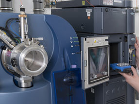
PerkinElmer Life Sciences Introduces KODAK MDS290 Microscopy Documentation System
Complete Solution for Light and Fluorescence Microscopy
BOSTON, MA - February 19, 2002 - PerkinElmer Life Sciences, a leading provider of drug discovery, Life Science research and genetic disease screening solutions, today announced the availability of the KODAK MDS290 for microscopy documentation that produces film-quality color images with the speed and convenience of digital imaging.
PerkinElmer Life Sciences customers will benefit from the versatility and efficiency of this new product offering. The KODAK MDS290 allows immediate access to captured microscopy images, while providing a full range of image enhancement and editing tools as it is seamlessly integrated with Adobe Photoshop software. The MDS290 maximizes PerkinElmer's Tyramide Signal Amplification (TSA)™ detection by combining color digital imaging with TSA's advanced signal amplification technology.
"Kodak's MDS290 is a complete microscopy imaging solution that broadens PerkinElmer's research imaging portfolio," said Paul Gillyon, vice president and general manager, Research Products, PerkinElmer Life Sciences. "PerkinElmer already offers a wide variety of imaging solutions for molecular biology research. However, this new easy-to-use offering further demonstrates our commitment to providing complete solutions to our customers."
With 2.1 megapixel resolution and color fidelity, the KODAK MDS290 provides publication-ready images of brightfield, darkfield and bright fluorescence specimens of up to 8" x 10."
Other news from the department research and development


Get the life science industry in your inbox
From now on, don't miss a thing: Our newsletter for biotechnology, pharma and life sciences brings you up to date every Tuesday and Thursday. The latest industry news, product highlights and innovations - compact and easy to understand in your inbox. Researched by us so you don't have to.
Most read news


More news from our other portals


See the theme worlds for related content
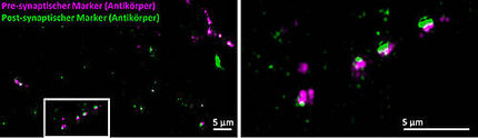
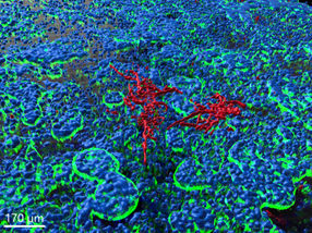
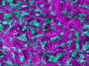
Topic world Fluorescence microscopy
Fluorescence microscopy has revolutionized life sciences, biotechnology and pharmaceuticals. With its ability to visualize specific molecules and structures in cells and tissues through fluorescent markers, it offers unique insights at the molecular and cellular level. With its high sensitivity and resolution, fluorescence microscopy facilitates the understanding of complex biological processes and drives innovation in therapy and diagnostics.