Cornell researchers make synthetic DNA 'barcodes' to tag pathogens, providing an inexpensive, off-the-shelf monitoring system
Advertisement


A supermarket checkout computer can identify thousands of different items by scanning the tiny barcode printed on the package. New technology developed at Cornell University could make it just as easy to identify genes, pathogens, illegal drugs and other chemicals of interest by tagging them with color-coded probes made out of synthetic tree-shaped DNA. A research group headed by Dan Luo, Cornell assistant professor of biological engineering, has created "nanobarcodes" that fluoresce under ultraviolet light in a combination of colors that can be read by a computer scanner or observed with a fluorescent light microscope.
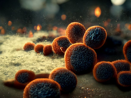
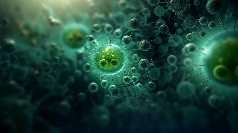
The researchers have tested their system using samples containing various combinations of E. coli, anthrax and tularemia bacteria and ebola and SARS viruses, and have found the color codes could clearly distinguish several different pathogens simultaneously. The idea is one of several applications the researchers have found for what they call "dendimer-like DNA," consisting of many short Y-shaped strands of DNA linked together in a treelike structure. Luo's research purposely and completely ignores the DNA's genetic coding properties. He uses DNA, he said, as a "generic instead of a genetic material." By synthesizing three short strands of DNA, each of which is complementary to one of the others along half its length, the researchers can create a Y-shaped structure. Combining several of these structures creates a web with many branching ends.
An antibody or some other molecule that will bind to the molecule to be detected is attached to one of the loose ends of the DNA. To other ends are attached molecules of fluorescent dye in a predetermined pattern. For example, one probe might contain four molecules of green dye and one of red. Another might have three molecules of green and two of red, and so on. If a mixture of several probes is added to a solution containing, for example, E. coli bacterial DNA, only probes with a particular color code will be programmed to bind to that DNA. The results can be seen under a fluorescent light microscope using colored filters that pass only one color at a time. A signal in which the ratio of intensity of green light is four times that of red light, for example, identifies a "4G1R" probe. The researchers say that up to 1,000 different codes can be created using only three fluorescent dyes.
To amplify the signals, the researchers attached many DNA probes to the surface of polystyrene microbeads 5.5 microns (millionths of a meter) in diameter. The results can be read in several ways. One is in a flow cytometer, in which samples move rapidly past a window where a computer reads the color codes of individual beads. Another is by dot blotting, in which the sample is spread on a sheet of absorbent paper and made visible to the naked eye. Or the color can be observed directly through a fluorescent light microscope, which is useful in situations where the geographic distribution of the target molecules is important, Luo said.
For convenience, a computer can convert the subtle differences in light intensity between, say 4G1R and 3G1R, into "pseudo colors," perhaps making one appear as orange and the other as pink, to make the difference clear to a human eye. The researchers point out that the nanobarcode detection system does not require complex preparation of a sample and can be applied to living cells. The technology could be used in genomic research, clinical diagnosis, drug testing, environmental monitoring and monitoring for biological terrorism, they suggest.
Original publication: Y. I. Luo, Y. T. H. Cu; "DNA fluorescence nanobarcodes"; Nature Biotechnology 2005.


Other news from the department science


These products might interest you


Most read news


More news from our other portals


See the theme worlds for related content
Topic World Cell Analysis
Cell analyse advanced method allows us to explore and understand cells in their many facets. From single cell analysis to flow cytometry and imaging technology, cell analysis provides us with valuable insights into the structure, function and interaction of cells. Whether in medicine, biological research or pharmacology, cell analysis is revolutionizing our understanding of disease, development and treatment options.