An advanced imaging technique detects cancer with unprecedented precision
New endoscopy technology enables early detection of esophageal cancer

Advertisement


Researchers from Helmholtz Munich, the Technical University of Munich (TUM), and the Medical University of Vienna have developed an advanced imaging technique called "O2E" that allows clinics to detect cancerous lesions in the esophagus with unprecedented precision. Published in Nature Biomedical Engineering, the study demonstrates that this innovative endoscopy technology reveals even the smallest pathological tissue changes, significantly improving early detection and diagnosis.
Previously Hidden Changes Now Visible
Esophageal cancer ranks among the deadliest cancers: when diagnosed at an advanced stage, the survival rate is only about ten percent. However, if detected early, around 90 percent of patients survive. The new O2E technology could play a crucial role in identifying changes in esophageal tissue at much earlier stages.
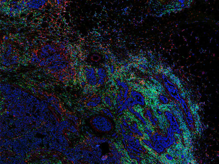
O2E combines two imaging techniques in a new endoscopy technology. While optical coherence tomography is particularly effective at capturing tissue structures, optoacoustic imaging – a method that stimulates tissue with light pulses and detects ultrasound signals resulting from the illumination – can visualize even the smallest blood vessels in deeper tissue layers. By merging these techniques, high-resolution 3D images of tissue structure and function in the esophagus are generated. Both sensors are integrated into an endoscopy capsule that scans the tissue in a full 360-degree angle.

“Our dual imaging system uncovers critical features of early cancer lesions, including microscopic structural changes beneath the mucosal surface and subtle microvascular alterations within the cancerous tissue, that previous methods were unable to detect,” says Prof. Vasilis Ntziachristos, Director at the Institute of Biological and Medical Imaging at Helmholtz Munich and Chair at TUM.
In their pilot study, the researchers examined the esophagus of animals and tissue samples from patients with Barrett’s esophagus, a precursor to esophageal cancer. They successfully identified distinct differences between healthy tissue, tissue with abnormal cellular changes, precancerous stages, and malignant tumors. Initial proof-of-principle tests were carried out on the inner lip of a volunteer, as it shares similar tissue characteristics with the esophagus.
Next Steps: Optimization for Clinical Application
Building on these promising results, a new EIC pathfinder project called ESOHISTO was granted and started in 2025. Funded by the European Union, EIC (European Innovation Council) Pathfinder projects support early-stage, high-risk research with the potential to drive transformative innovations. The researchers are now working to further refine the capsule technology to ensure it delivers high-quality imaging for use in humans.
“We also plan to integrate confocal endo-microscopy — a technique that provides high-resolution, real-time visualization of cellular structures — to enable more detailed analysis during examinations,” explains Dr. Qian Li, first author of the study from the Medical University Vienna. “This could pave the way for high-resolution endoscopic molecular imaging, allowing us to target specific molecular markers in cancer.” Ultimately, the researchers hope their approach will reduce the need for multiple biopsies and accelerate diagnostic processes in the future.
ESOHISTO will advance and validate the technology in preparation for its future market launch. In addition to the clear benefits for patients, the researchers believe that the healthcare system would also benefit from this early detection technology. While the treatment of advanced esophageal cancer typically costs around 140,000 euros per patient, early detection could reduce this cost to approximately 10,000 euros. Therefore, early detection does not only save the lives of many patients but will also result in substantial savings for the healthcare system.


Other news from the department science


