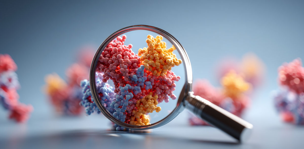
New method for protein analysis
The process can be used universally without the need for expensive special equipment
Advertisement


An international team led by the University Medical Center Hamburg-Eppendorf (UKE) has developed a new method for the high-resolution analysis of tissue samples. With the "Pathology-oriented MultiPlexing (PathoPlex)" method, more than 100 different proteins can be examined simultaneously in a single tissue sample. The method can be used universally without the need for expensive special equipment. The scientists hope that the new method could be used to identify disease-causing changes in the kidneys, for example, and enable personalized therapies. The researchers led by Prof. Dr. Victor Puelles from the III Medical Clinic and Polyclinic of the UKE, with the participation of the Institute for Medical Systems Biology, have published their study in the journal Nature.
Technological breakthrough with exceptional resolution
PathoPlex works in stepwise repeating cycles: First, different proteins are labeled with antibodies, then images are taken, then the antibodies are removed and the next cycle is started to stain further proteins. In the study, the researchers carried out a total of 95 such imaging cycles. Confocal microscopy was used to capture a total of 600 billion pixels with a pixel resolution of 80 nm - around 1000 times smaller than a human hair.
"A decisive advantage of PathoPlex is its compatibility with almost any fluorescence microscope - from simple wide-field microscopes to high-resolution confocal systems," says study leader and last author Prof. Puelles. The method uses commercially available antibodies and can process up to 40 archive samples in parallel. To cope with the large amounts of data, the researchers developed the "spatiomic" software, which automatically identifies protein co-expression patterns, enables spatial analyses and is freely available.
New insights into kidney diseases
When investigating diabetic kidney diseases with PathoPlex, the scientists identified stress signals in the renal tubules as new, potentially disease-causing effects. Changes could also be detected in type 2 diabetes patients without loss of kidney function - an important step towards improved early detection and treatment of diabetes complications. The method also proved to be suitable for evaluating the effect of antidiabetic drugs such as SGLT2 inhibitors in this patient group.
International collaboration
The multidisciplinary study was a collaboration between the UKE and the University of Aarhus (Denmark) as well as numerous international researchers in Japan, Germany, Australia, France, Switzerland and the USA. The project was funded by the German Research Foundation and the German Federal Ministry of Research, Technology and Space, among others.
Note: This article has been translated using a computer system without human intervention. LUMITOS offers these automatic translations to present a wider range of current news. Since this article has been translated with automatic translation, it is possible that it contains errors in vocabulary, syntax or grammar. The original article in German can be found here.
Original publication
Kuehl, Puelles et al. Pathology-oriented multiplexing enables integrative disease mapping. Nature. 2025.


Other news from the department science


These products might interest you


Most read news


More news from our other portals


See the theme worlds for related content
Topic world Protein analytics
Protein analytics provides a deep insight into these complex macromolecules, their structure, function and interactions. It is essential for discovering and developing biopharmaceuticals, understanding disease mechanisms, and identifying therapeutic targets. Techniques such as mass spectrometry, Western blot and immunoassays allow researchers to characterize proteins at the molecular level, determine their concentration and identify possible modifications.
Topic world Fluorescence microscopy
Fluorescence microscopy has revolutionized life sciences, biotechnology and pharmaceuticals. With its ability to visualize specific molecules and structures in cells and tissues through fluorescent markers, it offers unique insights at the molecular and cellular level. With its high sensitivity and resolution, fluorescence microscopy facilitates the understanding of complex biological processes and drives innovation in therapy and diagnostics.