3D bioprinting: improved nutrient transport in printed tissue
NMI, TU Darmstadt and Black Drop develop improved bioink
Advertisement


How can we 3D print tissues that mimic the complex anatomy of natural body tissue as closely as possible? 3D bioprinting is a great hope in the field of regenerative medicine to produce miniaturized tissues and organ precursors with biological functionality. Today, however, scientists are still working on the challenge of producing a printable and at the same time compatible starting material. Researchers at the NMI Natural and Medical Sciences Institute in Reutlingen, TU Darmstadt and Black Drop Biodrucker GmbH have developed a new type of bioink that improves the transport of nutrients in printed tissue, for example.
Bioprinting: many challenges, many opportunities
3D bioprinting, bioink, electrospinning: what is it anyway? 3D printing has now found its way into many areas of life and, above all, the economy. It is a process in which a three-dimensional object is printed using a special starting material. In 3D bio-printing, this starting material is bio-ink, which contains living cells and is combined with hydrogels and biological factors, for example, to print organic objects. In addition, electrospinning can be used to produce wafer-thin fibers.
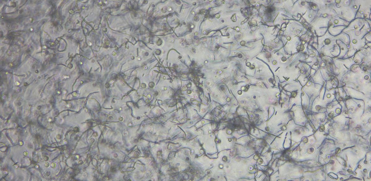
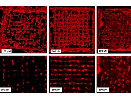
Electro-spun fibers enable nutrient transport
“With a diameter of 5-10 µm, these fibers are in the range of blood capillaries and are the significant advance in our bio-ink,” explains Dr. Hanna Hartmann, Head of Division at the NMI and inventor in the joint patent. Until now, the transport of nutrients in 3D-printed tissue has been a major problem. “The fibers now measurably improve this transport. The particularly exciting finding for us is that they don't even have to be hollow on the inside,” reports Jannik Stadler, site manager of Black Drop Biodrucker GmbH, which played a key role in the development of the bioink as coordinator of the BMBF-funded NatInk project. This bio-ink also has particularly advantageous properties in terms of its mechanical strength and swells less. Annabelle Neuhäusler, a doctoral student at the Institute for Printing Machines and Printing Processes at TU Darmstadt, was able to demonstrate this in the joint publication.
A step towards better tissue models for pharmaceutical research
In future, such improved bioinks could be used to produce tissue models for pharmaceutical research, for example. This could eliminate the need for animal testing and allow patient-specific active ingredients to be tested. Another field of application lies in the area of regenerative medicine. In addition to increasing nutrient diffusion, fiber integration helps to improve the mechanical properties of bioinks. This is particularly important for surgical applications, where handling, dimensional stability and primarily strength play a key role.


Other news from the department science


Most read news


More news from our other portals


Something is happening in the life science industry ...
This is what true pioneering spirit looks like: Plenty of innovative start-ups are bringing fresh ideas, lifeblood and entrepreneurial spirit to change tomorrow's world for the better. Immerse yourself in the world of these young companies and take the opportunity to get in touch with the founders.