Flexible neck in cell-receptor DC-SIGN targets more pathogens
Pathogen recognition is the foundation of the body's immune response and survival against infection. A small cell-receptor protein called DC-SIGN is part of the immune system, and recognizes certain pathogens, including those responsible for Ebola, dengue fever and HIV. How the molecule binds to pathogens has been unclear.
New findings from a research team led by University of Illinois chemist Deborah Leckband show that flexibility in the region near the binding sites of DC-SIGN plays a significant role in pathogen targeting and binding.
"Our work focuses on how DC-SIGN recognizes HIV and other pathogens, and on what structural features enable it to bind very tightly to those pathogens," said Leckband, the Reid T. Milner Professor of Chemistry at the U. of I. "Once we begin to understand the molecular design rules that lead to this tight binding, we can begin to design inhibitors to block this interaction."
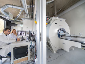
To study the binding behavior of DC-SIGN (Dendritic Cell-Specific Intercellular adhesion molecule-3-Grabbing Non-integrin), also known as CD209 (Cluster of Differentiation 209), the researchers used a device called a surface force apparatus.
The surface force apparatus measures the molecular forces between two surfaces as they are first brought together and then pulled apart. In the current work, the surfaces were cell receptor DC-SIGN and a target membrane decorated with carbohydrates to mimic a pathogen surface.
The forces were measured as a function of the distance between the two surfaces, which was measured with single-angstrom resolution.
"Our force-distance measurements provided the first direct, dynamic evidence for flexibility in the neck of DC-SIGN, and its possible role in pathogen recognition and binding," said Leckband, corresponding author of a paper accepted for publication in the Proceedings of the National Academy of Sciences, and posted on the journal's Web site.
From their force-distance measurements the researchers determined DC-SIGN's neck length as 28 nanometers, in agreement with hydrodynamic measurements and theoretical estimates by other researchers, which placed the neck length between 20 and 30 nanometers.
When the protein binds to a pathogen, binding sites on the cell receptor rearrange slightly, to adapt to the target surface and maximize the bond. This 5 nanometer conformational change is binding-induced, and made possible by a flexible linker in the neck, the researchers report.
"The protein neck region acts as a stiff, but flexible, rod that projects the molecule's binding sites away from the cell surface," Leckband said. "A rigid presentation of the binding sites at the end of the neck would restrict DC-SIGN to a few specific, spatial forms. Instead, the molecule's flexibility and adaptability allow it to recognize a much wider range of pathogens."
Other news from the department science


Get the life science industry in your inbox
From now on, don't miss a thing: Our newsletter for biotechnology, pharma and life sciences brings you up to date every Tuesday and Thursday. The latest industry news, product highlights and innovations - compact and easy to understand in your inbox. Researched by us so you don't have to.