Customized therapy for breast cancer patients
Researchers in the VPH-PRISM project develop new procedures to improve treatment of breast cancer using multidisciplinary image data
Advertisement


Every tenth woman is afflicted with breast cancer during her lifetime.The diagnosis and treatment of a patient involves the collaboration of a wide range of specialists. The broad range of software platforms for storage and visualization of medical imaging data used by radiologists, pathologists, oncologists, radiotherapists and surgeons, brings challenges for communication and data transfer between the platforms. Improved extraction and transfer of information between the platforms may support breast cancer diagnosis and therapy planning.
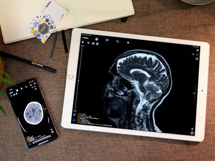
This is where the VPH-PRISM project comes into play. Eight research partners and one coordinating partner are developing novel software systems that intelligently connect medical data sets to allow innovative assistive functions. The researchers plan to combine image data generated by various diagnostic procedures (X-ray, MRI, tissue histology) for display in a single software application. Their vision consists of systematically gathering and quantitatively combining the image information currently available from various disciplines. For the first time, this would permit a large number of microscopic and macroscopic tissue parameters to be correlated efficiently and precisely. This could provide important indicators, enabling clinicians to customize therapy planning for individual patients.
To realize this, researchers in the project intend to create an interactive database to connect images with other relevant information, such as a patient’s risk of hereditary disease and environmental factors. This database would allow development of more focused therapy for breast cancer patients in the future. The Fraunhofer Institute for Medical Image Computing MEVIS in Bremen has taken charge of the scientific coordination of the project and is responsible for developing significant sections of the required image analysis procedures.
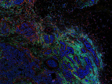
Early detection and therapy for breast cancer typically involves multiple disciplines. Radiologists interpret X-ray and MRI scans of the patient breast, whereas pathologists use 40x magnification and higher to examine the spatial arrangement of single cells from breast tissue samples. Surgeons attempt to remove malignant tumors effectively while conserving as much healthy tissue as possible. Radiotherapists treat breast cancer with radiation, and oncologists decide which type of chemotherapy is expected to be most beneficial.
An important challenge is to increase the interconnectivity of the information technology systems of these different disciplines. Radiologists and pathologists often work in the same hospital with different IT systems, each partly optimized to display and manipulate only the specialists’ own image data. When experts from different fields discuss suitable therapies, different image storage systems may result in abbreviated data transfer possibilities. Interdisciplinary tumor board meetings, during which specialists discuss individual breast cancer cases, may benefit from increased interconnectivity of the different systems.
To improve this situation, VPH-PRISM partners are developing software for X-ray, MRI images, ultrasound, and histology from biopsies, to provide a unified display to spatially superimpose, measure, and manipulate these images. Especially helpful would be the ability to characterize the area surrounding a tumor more accurately. The project will aim to answer the questions: has this tissue changed to such a degree that it must be removed with the tumor during the operation, or can a more local excision, exposing the patient to a less invasive operation, be performed without the risk of recurrence?
To accomplish this, tissue sample pathology slides must be digitized. However, digitization generates large amounts of data, which pathologists can only partially inspect. To solve this, VPH-PRISM experts are also developing software that automatically preselects and preprocesses the data, thereby facilitating the work of the pathologist. If the venture is successful, experts estimate that digital pathology will promote a breakthrough in breast cancer care similar to the effect that the widespread introduction of mammography had on early detection that has occurred over the last 15 years.
The project will explore how a deep understanding of tissue microstructure, gleaned from histology, can aid interpretation of X-ray, MRI, and ultrasound images. Additionally challenging is the presentation of tissue sample images alongside MRI and X-ray images. Experts using computationally intensive algorithms must guarantee that multiple data sets fit perfectly together. Only in this manner can tissue parameters gathered using different scalings be spatially correlated and hence superimposed.
The goal is a software tool that supports clinicians when choosing therapy. Patient data should be grouped automatically according to shared criteria. This allows the software to provide clues for optimal chemotherapy, for instance, if a patient with particular tissue characteristics has been assigned to a particular group. This could prove useful for monitoring the progress of therapy more accurately: Is the tumor degenerating as quickly as expected when a patient undergoes a certain type of chemotherapy? If not, then the doctor could cease ineffective therapy in a timely fashion and provide the patient with alternative drugs.


Other news from the department science


These products might interest you