Detection of cancer biomarkers from blood samples using nanopore-based DNA computing technology
Advertisement


cholangiocarcinoma, also known as bile duct cancer, is a cancer type with a characteristically high mortality. At the time of diagnosis, most bile duct cancers are typically already incurable. This is why methods for the early diagnosis of bile duct cancer are urgently needed.
Pattern recognition of miRNA expression using DNA computing and nanopore decoding
Ryuji Kawano, Tokyo University of Agriculture and Technology
Liquid biopsy, the sampling of non-solid biological tissue like blood, is gaining interest as a quick and non-invasive method for diagnosing cancers. Unlike traditional biopsies that require surgery and often general anesthesia, a liquid blood biopsy only requires a couple of milliliters of blood, with minimal harm to the patient.
After sampling, the blood is screened for specific markers indicating the presence of cancerous tissue. For example, specific patterns of microRNA (miRNA), short non-coding strands of RNA, are associated with different types of cancer and can be used to diagnose cancers from liquid biopsies with high precision. However, the low concentration of miRNA in the blood samples makes their detection challenging.
Researchers at the Tokyo University of Agriculture and Technology have developed a new method for the detection of cancer miRNA patterns based on DNA computing technology. The developed method shows potential as a promising tool for simple and early cancer diagnosis from liquid biopsies with low concentrations of target biomarkers. The findings were published in the peer-reviewed journal JACS Au on June 26th, 2022.
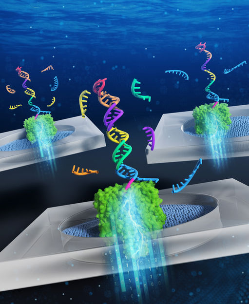
“DNA computing uses the biochemical reactions of the information-encoding DNA molecules to solve problems based on formal logic, in the same way that normal computers do,” said corresponding author Ryuji Kawano, professor in Tokyo University of Agriculture and Technology (TUAT) in Japan. “In this case, a diagnostic DNA molecule was designed to be able to bind five different kinds of miRNA associated with bile duct cancer. In the process of binding the miRNA molecules, the diagnostic DNA converts the expression pattern of the miRNAs into the information contained in the form of a nucleic acid structure.”
To read this information, the scientists use a method called nanopore decoding. In this method, the DNA is passed through a nano-sized hole, or “pore”. As the molecule transits the pore, it will obstruct the flow of electrical current through the pore. These perturbations in the current through the pore can be then measured and used to deduce the properties of the passing molecule. In the case of the diagnostic DNA, the bound miRNAs will be “unzipped” from the DNA, resulting in a current inhibition of characteristic amplitude and duration. Through statistical analysis of the unzipping data of the miRNA patterns, the scientists were able to recognize cancer-specific expression patterns even from clinical samples with extremely low concentrations of miRNA. This is a significant improvement in the field of nanopore diagnosis, as nanopore measurements have generally been considered unable to detect nucleic acids at such low levels, which has undermined the use of the technology in clinical applications.
Original publication


Other news from the department science