Fluorescent molecules reveal how cancer cells are inhibited
Advertisement
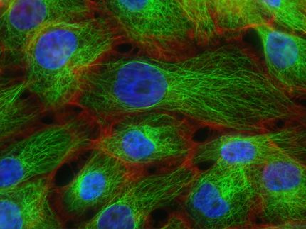
A team of researchers at Lund University in Sweden has developed a fluorescent variant of a molecule that inhibits cancer stem cells. Capturing images of when the molecule enters a cell has enabled the researchers, using cell-biological methods, to successfully describe how and where the molecule counteracts the cancer stem cells.
Salinomycin is a molecule produced by terrestrial bacteria of the species Streptomyces albus. It was previously known that this molecule acts selectively against cancer stem cells, but the mechanism behind it was not understood. Now, Lund researchers have created a fluorescent variant of salinomycin to understand how it works.
"We have shown where the molecule ends up when it is absorbed by cancer cells. By making the molecule fluorescent, we have also been able to capture the course of events on film", says Daniel Strand who leads an organic chemistry research team at Lund University.
It has long been known that this molecule can transport ions across cell membranes, in this case potassium ions. Even so, the researchers were surprised when they saw images of the molecule in cells.
"Those of us involved in the study initially naïvely assumed that the molecule acted in the cell's outer membrane", says Daniel Strand.
However, the images showed that the molecule rapidly passed through the outer cell membrane and its destination was an organelle called the endoplasmic reticulum. This is where the molecule acts as an ion transporter, and it is this specific activity that the researchers have succeeded in connecting to a reduction in the percentage of cancer stem cells.
The research results may contribute new approaches to the development of cancer drugs both for treatment of cancer and for reducing the risk of relapse.
Original publication
Xiaoli Huang, Björn Borgström, John Stegmayr, Yasmin Abassi, Monika Kruszyk, Hakon Leffler, Lo Persson, Sebastian Albinsson, Ramin Massoumi, Ivan G. Scheblykin, Cecilia Hegardt, Stina Oredsson, and Daniel Strand; "The Molecular Basis for Inhibition of Stemlike Cancer Cells by Salinomycin"; ACS Central Science; 2018


