New microfluidic chip can help identify unwanted particles in water and food
Advertisement


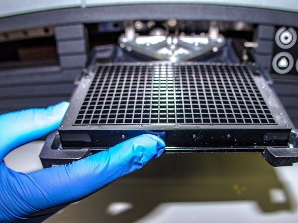
A new process for making a three-dimensional microstructure that can be used in the analysis of cells could prove useful in counterterrorism measures and in water and food safety concerns.
The research, conducted by members of Virginia Tech's Microelectromechanical Systems Laboratory (MEMS) Laboratory in the Bradley Department of Electrical and Computer Engineering, is the focus of a recent article in the Institute of Electrical and Electronic Engineers' Journal of Microelectomechanical Systems.
In their engineering laboratory, the researchers developed a new microfabrication technique to develop three-dimensional microfluidic devices in polymers. Microfluidics deals with the performance, control, and treatment of fluids that are constrained in some fashion, explained Masoud Agah , director of the laboratory.

As a result of this work, Agah, associate professor of the Bradley Department of Electrical and Computer Engineering and of the Virginia Tech–Wake Forest School of Biomedical Engineering and Sciences, and Amy Pruden, professor of civil and environmental engineering at Virginia Tech, have received a National Science Foundation award of $353,091 to use the technology and develop new microchips named 3D-πDEP standing for "three-dimensional, passivated-electrode, insulator-based dielectrophoresis" for pathogen detection.
The NSF grant will allow them to focus on the isolation of waterborne pathogens that represent one of the "grand challenges to human health, costing the lives of about 2.5 million people worldwide each year," Agah and Pruden said.
According to the World Health Organization, the isolation of pathogenic bacteria from the environment has not significantly changed since the 1960s, when methods for chemical treatment of samples to remove background organisms were first implemented.
In the past, Agah said, researchers have mainly used two-dimensional microfluidic structures since this type of fabrication is more simplistic. With the three-dimensional device developed by Agah and his collaborators, Yayha Hosseini and Phillip Zellner, both graduate students in the department, they are able to customize the shapes of the channels and cavities of the devices the fluids passed through.
The advantage of the fabrication process is that with a very economical technique it creates three-dimensional varying channels and cavities in a microfluidic structure with rounded corners as well as many other customized shapes.
These shapes are important because they resemble the living conditions as they occur naturally and this allows the use of the three-dimensional microfabrication technology beyond pathogen detection.
As an example, in human blood vessels, cells interact with each other and their surrounding environment inside circular channels. They have varying diameters, along with multiple branching and joints.
"Only under this type of condition can one truly study the biology of cells within a system in vitro as if it is occurring in vivo – our new microfluidic fabrication technology can resemble more realistically the structures of a cell's true living conditions," Agah said. It is the introduction of the three-dimensions that provides this distinctive environment.
To make their three-dimensional structure, the Virginia Tech researchers used the material polydimethylsixolane, known for its elastic properties similar to rubber. This material is already widely used because of its transparency, biocompatibility, and low-cost.
"Our work establishes a reliable and robust, yet low-cost technique for the fabrication of versatile 3-D structures in polydimethylsixolane," Agad said.
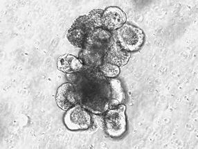
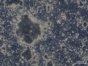
Microfluidic devices can be used to trap and sort living organisms such as bacteria, viruses, and cells. With this new three-dimensional device that has a higher sensitivity and throughput than the two-dimensional version, according to Agah, he is able to make their predictions of applications ranging from water and food safety to fighting biological and chemical terrorism and to healthcare by fishing for abnormal cells in body fluids.
Other news from the department science