Investigating kinase activity in living cells: Scientists build molecular recording tool
Overcoming limitations of optical imaging

Advertisement


The ability of protein kinases to transfer a phosphate group to target proteins plays an important role in many cellular processes. Scientists at the Max Planck Institute for Medical Research in Heidelberg have now developed a novel molecular tool that can monitor these kinase activities both spatially and temporally. This makes it possible to investigate the link between kinase activities and cellular phenotypes in heterogenous cell populations and in vivo.
Protein kinases, an important subgroup of kinases, control most cellular processes. Their activity is crucial for regular cell processes, whereas aberrant kinase activity is involved in numerous diseases.
Overcoming limitations of optical imaging
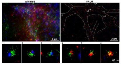
“We wanted to find a way to record kinase activities in a scalable and high-resolution manner, without the restrictions of optical imaging. Under the microscope, observations are typically limited to real-time imaging of a relatively small number of cells and are not readily applicable to deep tissues”, says De-en Sun, who led the project team together with Kai Johnsson, director at the Max Planck Institute (MPI) for Medical Research.
Key: converting transient kinase activity into a stable fluorescent signal
With their molecular recorder Kinprola, the Max Planck scientists found a way to overcome the limitations of real-time optical recording. They make use of a molecular switch depending on phosphorylation, a process that is catalyzed by kinases. When this kinase is activated, recording is initiated by washing in a fluorescent substrate, which enables labeling of activated Kinprola proteins. The recording is terminated by washing out the substrate. The labeled Kinprola population remains stable over time, while the rest of the Kinprola protein pool remains unlabeled.
This procedure enables decoupled recording and analysis, using different imaging techniques and flow cytometry – a technique that enables the physical and chemical properties of cells or other particles to be analyzed and sorted –, depending on the experiment. It converts transient kinase activity into a stable fluorescent signal, and is characterized by its high versatility and scalability. Kinprola works for multiple kinases, and functions in vivo. In mouse brains, for instance, Kinprola can record PKA activation elevated by drug injection.
Cooperation partners from the Heidelberg scientific community and China
De-en Sun from the MPI for Medical Research in Heidelberg is supported by the Humboldt Research Fellowship and an interinstitutional postdoctoral fellowship of the Health + Life Science Alliance Heidelberg Mannheim. Scientists at the German Cancer Research Center (DKFZ) and Heidelberg University contributed to the transcriptomics (Frank Winkler and Wolfgang Wick groups) and CRISPR screening (Michael Boutros group). Scientists of the Yulong Li group at the Peking University, China, contributed to the mouse experiments.
Original publication
De-en Sun, Siu Wang Ng, Yu Zheng, Shu Xie, Niklas Schwan, Paula Breuer, Dirk C. Hoffmann, Julius Michel, Daniel D. Azorin, Kim E. Boonekamp, Frank Winkler, Wolfgang Wick, Michael Boutros, Yulong Li, Kai Johnsson; "Molecular recording of cellular protein kinase activity with chemical labeling"; Nature Chemical Biology, 2025-7-10


Other news from the department science


Most read news


More news from our other portals


See the theme worlds for related content
Topic World Cell Analysis
Cell analyse advanced method allows us to explore and understand cells in their many facets. From single cell analysis to flow cytometry and imaging technology, cell analysis provides us with valuable insights into the structure, function and interaction of cells. Whether in medicine, biological research or pharmacology, cell analysis is revolutionizing our understanding of disease, development and treatment options.