How rogue communications between cells lead to Leukaemia
How do cells communicate in blood formation?
Advertisement


How do biochemical messengers mediate the development of new blood cells and how do these processes get out of control in leukaemias? An international research team involving partners from Germany, United Kingdom, Finland and the USA has achieved a fundamental breakthrough in understanding the mechanism of these processes.
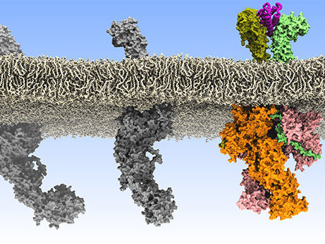
University of Helsinki/ Ilpo Vattulainen and Joni Vuorio
In adults, billions of mature blood cells are formed from haematopoietic stem cells in the bone marrow every day. This process is tightly regulated by a family of messenger proteins called cytokines that control the development and proliferation of the different blood cell types. Cytokines interact with specific receptors on the surface of cells, which allow the transmission of signals controlling whether the cell divides, or differentiates into a specific blood cell type. Various leukaemias are associated with genetic mutations that activate these signalling pathways in the absence of cytokines in an uncontrolled manner. Until now, the molecular mechanisms of how individual mutations trigger signal activation and lead to these blood cancers have remained unclear.
First author Dr. Stephan Wilmes, who started the project as a Postdoc at Osnabrück University: “It was truly inspiring to tackle this highly relevant biomedical question by applying cutting-edge biophysical techniques.” “A particular challenge was to coordinate the different research approaches of the participating working groups," says Maximillian Hafer, who has since assumed responsibility for the project.
Using single-molecule microscopy in living cells, the researchers have now been able to clearly show for the first time that the receptors are crosslinked by cytokines to form pairs. Until now, it has been assumed that the receptors are already present as inactive pairs even without cytokines. From their new observations using super-resolution fluorescence microscopes, the researchers concluded that pair formation itself is the basic switch for the activation of signal transduction in the cell. “By directly visualising individual receptors at physiological conditions under the microscope, we were able to resolve a controversy that has preoccupied the field for more than 20 years," explains Professor Jacob Piehler from Osnabrück University.
In combination with biomedical studies at the Universities of York and Dundee, the researchers found that several important disease-relevant mutations led to the pairing of certain receptors without cytokine. "These observations led us to a previously unknown mechanism how individual mutations at this receptor trigger cytokine-independent signalling and thus can promote leukaemia," reveals Professor Ian Hitchcock from the University of York. Cooperation partners at the University of Helsinki used these insights to develop a comprehensive structural model via atomic-scale simulations and molecular modelling, which could explain the different modes of action of different mutations. "Our biomolecular simulations unveiled surprising features concerning the orientation of active receptor pairs at the plasma membrane, explaining how mutations render activation possible without a ligand. These predictions were subsequently confirmed experimentally", explains Professor Ilpo Vattulainen from the University of Helsinki.
These fundamental insights into the mechanism of signal activation enable completely new and much more targeted strategies for combating leukaemias. Further, the researchers suspect that a wide range of inflammatory and allergic diseases can also be traced back to similar mechanisms.
Original publication


Other news from the department science


Most read news


More news from our other portals


See the theme worlds for related content
Topic world Fluorescence microscopy
Fluorescence microscopy has revolutionized life sciences, biotechnology and pharmaceuticals. With its ability to visualize specific molecules and structures in cells and tissues through fluorescent markers, it offers unique insights at the molecular and cellular level. With its high sensitivity and resolution, fluorescence microscopy facilitates the understanding of complex biological processes and drives innovation in therapy and diagnostics.