DNA-guided 3-D printing of human tissue is unveiled
Technique produces 'organoids' useful in cancer research, drug screening
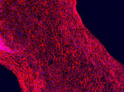
A UCSF-led team has developed a technique to build tiny models of human tissues, called organoids, more precisely than ever before using a process that turns human cells into a biological equivalent of LEGO bricks. These mini-tissues in a dish can be used to study how particular structural features of tissue affect normal growth or go awry in cancer. They could be used for therapeutic drug screening and to help teach researchers how to grow whole human organs.
The new technique - called DNA Programmed Assembly of Cells (DPAC) - allows researchers to create arrays of thousands of custom-designed organoids, such as models of human mammary glands containing several hundred cells each, which can be built in a matter of hours.
There are few limits to the tissues this technology can mimic, said Zev Gartner, PhD an associate professor of pharmaceutical chemistry at UCSF. "We can take any cell type we want and program just where it goes. We can precisely control who's talking to whom and who's touching whom at the earliest stages. The cells then follow these initially programmed spatial cues to interact, move around, and develop into tissues over time."
"One potential application," Gartner said, "would be that within the next couple of years, we could be taking samples of different components of a cancer patient's mammary gland and building a model of their tissue to use as a personalized drug screening platform. Another is to use the rules of tissue growth we learn with these models to one day grow complete organs."
But studying how the cells of complex tissues like the mammary gland self-organize, make decisions as groups, and break down in disease has been a challenge to researchers. The living organism is often too complex to identify the specific causes of a particular cellular behavior. On the other hand, cells in a dish lack the critical element of realistic 3-D structure.
"This technique lets us produce simple components of tissue in a dish that we can easily study and manipulate," said Michael Todhunter, PhD, who led the new study with Noel Jee, PhD, when both were graduate students in the Gartner research group. "It lets us ask questions about complex human tissues without needing to do experiments on humans."
To specify the 3-D structure of their organoids, Gartner's team makes use of a familiar molecule: DNA. The researchers incubate cells with tiny snippets of single-stranded DNA engineered to slip into the cells' outer membranes, covering each cell like the hairs on a tennis ball. These DNA strands act both as a sort of molecular Velcro and as a bar code that specifies where each cell belongs within the organoid. When two cells incubated with complementary DNA strands come in contact, they stick fast. If the DNA sequences don't match, the cells float on by. Cells can be incubated with several sets of DNA bar codes to specify multiple allowable partners.
To turn these cellular LEGOs into arrays of organoids that can be used for research, Gartner's team lays down the cells in layers, with multiple sets of cells designed to stick to particular partners. Not only does this let them build up complex tissue components like the mammary gland, but also to experiment with specifically adding in a single cell with a known cancer mutation to different parts of the organoid to observe its effects.
To demonstrate the precision of the technique and its ability to generalize to many different human tissue types, the research team created several proof-of-principle organoid arrays mimicking human tissues such as branching vasculature and mammary glands.
Original publication
Other news from the department science


Get the life science industry in your inbox
From now on, don't miss a thing: Our newsletter for biotechnology, pharma and life sciences brings you up to date every Tuesday and Thursday. The latest industry news, product highlights and innovations - compact and easy to understand in your inbox. Researched by us so you don't have to.